Cubital Tunnel Syndrome

Hand Health Resources considers Cubital Tunnel Syndrome as the second most common nerve entrapment syndrome occurring in the upper body or arm. Increased compression or pressure on the ulnar nerve in the elbow gives rise to this condition. Along with discomfort, this pathology may lead to the loss of hand functioning.

Cubital Tunnel Syndrome

What do you understand by Cubital Tunnel Syndrome?

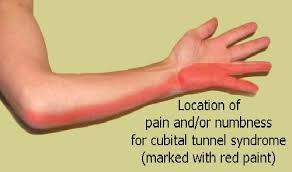
Ulnar nerve is a crucial nerve running through your arm and is responsible for sensation and movement of the forearm and hand as well. This Ulnar nerve passes through a tunnel of soft tissues named as ‘cubital tunnel’ that runs under a bony bump at the inner side of your elbow. The syndrome is called ‘cubital tunnel syndrome’ as it results due to the entrapment or irritation of nerve within the confines of cubital tunnel. This condition affects more men than women and the athletic performance of the athletes who are engaged in sports requiring strong hand or wrist movements. The entrapment causes tingling, numbness and pain along the inside of your arm down the ring and little finger.

What causes Cubital Tunnel Syndrome?

Usually, the syndrome occurs when the Ulnar nerve within the constricted enclosure of cubital tunnel is stretched compressed or irritated as there is a very little soft tissue to protect it. Other factors leading to the Cubital Tunnel Syndrome may include:

  • Keeping the elbow bent for longer periods or repeated bending of the elbow
  • Back and forth sliding of the nerve over the medial epicondyle
  • Leaning on your elbow for longer periods of time
  • Fluid build-up in the elbow
  • A direct blow to the inside of the elbow as in the case of sports like; baseball, tennis, rugby, football etc.
  • Athletic activities including repetitive overhead motions like; javelin throw, tennis serve or baseball
  • Failure to warm up  and stretch properly before a sport
  • Elbow fractures, bone spurs, trauma or cysts

What are the treatment options suggested to treat Cubital Tunnel Syndrome?

Cubital Tunnel Syndrome usually responds to Physical Therapy. Initially the therapists advise to modify and avoid the activities, postures and positions causing Cubital Tunnel Syndrome. Other treatment options may include:

  • Therapists will advice to avoid repeated flexion, over leaning and pressurizing the elbow
  • Gentle exercises may be prescribed to ease the ulnar nerve glide through Cubital Tunnel
  • therapist may recommend or customize an elbow splint to keep your arm straight
  • Stretching exercises may be prescribed to achieve full range of motion
  • Therapist may provide a home exercise program and educate you about proper body mechanics
  • Strengthening exercises like; strengthening of muscle groups, bending and straightening of elbow, forearm movement may be prescribed
  • Nerve Gliding exercises may be advised to help the arm and wrist from getting stiff
  • Range of motion exercises may be prescribed to enhance the mobility without causing pain or discomfort
  • Special exercises to help the Ulnar glide and the exercises that mimic your daily and work activities may be prescribed

Contact Alliance Physical Therapy for the state of the art treatment of any of your musculoskeletal injury or problem. Our certified therapists use patient proven methods and develop customize treatment plans according to your needs and urgencies.
 

Alliance Physical Therapy Clinics offer Treatments for Auto- Accident Injuries


Auto Injuries Rehab

Auto-Accidents are said to happen every 5 seconds in the United States and these figures continue to rise. Victims of auto-accidents usually suffer from skeletal, muscular, neurological and soft-tissue injuries which are not detectable initially as they do not cause much pain. So the victims ignore them and do not seek professional treatment. This causes the biggest blunder. With auto-accident injuries, immediate and specialized treatment is needed to avoid developing long term discomfort and chronic pain. A pro-active Physical Therapy treatment and Rehabilitation plan can save the victims from surgical interventions and long term reliance on medications as well.

Physical Therapy modalities while conditioning your muscles and bones also help to alleviate pain and relieve the debilitating effects of auto-accident injuries. Vehicular injuries often develop the symptoms like; back pain, whiplash, fatigue, headache, muscle-spasms, nausea, neck pain, tendinitis, shoulder pain, problems with vision, dizziness etc. Physical Therapists are experts at diagnosing, evaluating and treating these injuries. They re-align your vertebrae and help to restore your lost structural balance and treat soft tissue injuries with customized rehabilitation program.

Various structural evaluation techniques including; X-rays, Spinal Joint Movement Evaluation, Gait and Postural Analysis, Joint Range-of Motion Testing, Strength and Reflex Evaluation, CAT or CT scan and MRI are conducted and adopted to detect and correct various musculoskeletal injuries.  Physical Therapy is mainly directed towards maximizing the motion of spine, curing spinal disc, alleviating muscle spasm and improvising the muscular strength. Following Physical Therapy modalities are used to treat auto-accident injuries:

  • Hands-On Therapy and therapeutic equipments are used for the vestibulo-ocular rehabilitation of the victims.
  • Corrective exercises are used to stretch and strengthen the damaged muscles.
  • Range-of Motion exercises are prescribed to improvise function and mobility
  • Spinal Decompression therapy is employed to relieve back pain due to herniated disc or Sciatica
  • Therapeutic exercises targeting and strengthening the joints are suggested.

So if you have been a victim of vehicular accident and sustained a musculoskeletal injury and want to get relief without pain-killers and surgical interventions, then Physical Therapy is the most influential treatment available.

Contact Alliance Physical Therapy for the skilled and prompt treatment of auto-accident injuries. Our board certified therapists are specialized in diagnosing, treating and managing auto-accident whiplash injuries and have helped numerous patients to recover successfully from whiplash.

Knobby Knees

Osgood Schlatter Disease

Osgood Schlatter Disease (OSD) or Knobby Knees is a common cause of knee-pain among the teenagers. This pain mostly affects the boys between 13 to 14 years of age and the girls between 11 to 12 years. This problem mostly occurs during the growth spurt.

What do we understand by Osgood Schlatter Disease?

Osgood Schlatter Disease is an overuse injury of the knee resulting in pain and swelling below the knee cap over the shin bone. This disease is marked by the inflammation of the bone cartilage and /or tendon at the top of the shin bone, where the tendon from the knee cap attaches. It commonly affects the teens who are engaged in vigorous sports activities which involve lots of running, jumping and swift direction changes, as in; basket ball, gymnastics, soccer, figure skating, ballet and volley ball.

What are the potential signs and symptoms of Osgood Schlatter Disease?

Osgood Schlatter Disease usually harms one knee at a time but it may affect the both knees. It lasts for months and recurs until the child stops growing. Following signs and symptoms may be observed in the teens with OSD:

  • Pain below the knee cap
  • Severe pain during and after the activity
  • A tiny and soft bony bump under the knee cap
  • The swelled area becomes tender to pressure
  • Limping after the physical activity
  • Pain Easing with Rest
  • Tightness in the muscles surrounding the knee.

What are the factors leading to the Osgood Schlatter Disease?

At times, Osgood Schlatter Disease develops for no apparent reason. But the below mentioned factors may lead to the condition:

  • Repeated small injuries to knee before the growth spurt is over
  • Overuse of thigh muscles resulting in swelling
  • Repeated strain on the attachment of the patellar ligament
  • Formation of callus (healing bone) resulting into hard bony bump

What are the treatment options suggested to heal Osgood Schlatter Disease?

Osgood Schlatter Disease usually heals itself after the child’s bones stop growing. Till then, a customized physical therapy treatment program targeted at relieving pain and swelling are practiced, following techniques are employed to treat the symptoms:

  • Activity modification and in more severe cases, activity elimination is advised
  • R.I.C.E. Therapy (Rest, Ice, Compression and Elevation) is suggested to relieve pain
  • Stretching exercises; concentrated on the stretching of thigh muscles, are recommended
  • Strengthening exercises may be advised to stabilize the knee joint
  • To shield the sensitive area, the physical therapists may recommend to wear knee pads during the sport
  • Bracing, strapping and wrapping of knee area may be recommended for support

Contact Alliance Physical Therapy

Contact Alliance Physical Therapy to proficiently manage and relieve any type of musculoskeletal pain. Our diversified team of trained and certified therapists will develop a customized treatment plan for you while taking due care of your needs and urgencies and will help you to regain health and proper functioning.

Becker Muscular Dystrophy

Becker Muscular Dystrophy Treatment

What do we mean by Becker Muscular Dystrophy?

Muscular Dystrophy belongs to that group of genetic degenerative disorders which involve progressive weakness and wasting of voluntary muscles of the body. This condition is named after the German doctor Peter Emil Becker who first described the disorder.

Becker Muscular Dystrophy (BMD) is among those common nine kinds of dystrophies; which are characterized by the slow weakness and wasting of skeletal muscles that are used for movement and heart muscles as well. Such dystrophies are common among the men than women. Being a genetic disorder; your family history of disease increases your risk of getting affected.

Despite of being more common in men than women; males usually inherit it from their mothers, i.e. females are the carriers of this disease but they usually do not exhibit any symptoms.

How is the Becker Muscular Dystrophy an inherited disease?

When a particular flawed gene or mutation of a particular gene on the X-chromosome makes its associated protein (named as dystrophin) partially functional, this results in the Becker Muscular Dystrophy. That is the reason that Becker Muscular Disease is also known as ‘X-linked recessive genetic defect’ i.e. it is inherited due to a defective gene on the X-chromosome.

Males manifest the symptoms of this disease because they have only one X- chromosome. While the females have two X- chromosomes, so they usually do not show any signs or symptoms as the healthier X- chromosome compensate for the defective one.

What are the signs and symptoms of Becker Muscular Disease?

The symptoms usually begin to appear during the childhood and are diagnosed by the age of 11 years. But the severity of the disease varies from person to person. The symptoms may include:

• Gradual but progressive muscle weakness
• Muscles cramps on exercising
• Inability to walk independently by the age of 16
• Loss of walking by the age of 40 -60 and at times, 20-30
• Severe upper extremity and trunk muscle weakness
• Toe-Walking and frequent falls
• Difficulty in breathing and Heart disease; particularly, Dilated Cardiomyopathy
• Skeletal and Muscular Deformities
• Fatigue
• Difficulty in getting up from the floor and Climbing stairs
• Loss of balance, co-ordination and muscle mass

What are the treatment options suggested to treat Becker Muscular Dystrophy?

The treatment is generally focused at treating the symptoms and enhancing the patient’s quality of life. Treatment options suggested are generally supportive and include the following:
• Physical therapy is suggested to encourage the activity as the inactivity (like bed rest) can further worsen the condition. Muscle strengthening exercises are suggested to maintain muscle strength.
• Massage, Compression treatment and night Splints may be advised to treat muscle cramps.
• Use of assistive devices like; knee, leg or back braces are suggested to keep the muscles flexible.
• Occupational Therapy is advised; where patients are helped and taught to use orthopedic appliances like; wheel chairs and other assistive devices as well to improve movement and training for doing the daily tasks in new ways is also provided.

Contact Alliance Physical Therapy for the adequate and enhanced treatment of any of your neuromuscular disorder. Our certified therapists develop customized treatment plans according to your needs and preferences and utilize patient proven treatment techniques to heighten the quality of your life.

Anterior Knee Pain

Chondromalacia Patellae: Causes, Symptoms and Treatment


Chondromalacia Patellae

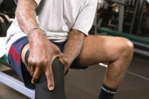
Chondromalacia Patellae, also known as ‘runner’s knee’ is the most common cause of chronic knee pain. It often affects the young athleticsbut may also happen to-older people with arthritis and females as well, as they have less muscle mass than males.

What do we mean by Chondromalacia Patellae?

Chondromalacia Patellae is a condition, where the cartilage under your patella or knee cap softens and wears away or deteriorates. Poor alignment or overuse of knee cap or patella wears down the cartilage and as a result, knee cap begins to rub against the thigh bone which further leads to grinding sensations and chronic pain in the front of the knee.

What are factors leading to Chondromalacia Patellae?

When you bend your knee; the backside of your knee cap slides over the femur (thigh bone). But when the knee cap does not move properly it begins to rub with the thigh bone leading to swelling and pain. Many factors including the following lead to the improper movement of knee cap which ultimately results in Chondromalacia Patellae:

  • Weak hamstrings and quadriceps (back and front thigh muscles)
  • Imbalance between the adductors and abductors (the muscles on the outside and inside of your thigh)
  • Improper tracking of knee cap
  • Trauma to knee cap like; Dislocation or Fracture
  •  Repetitive stress on knee joint due to activities like; running, jumping, twisting, skiing and playing soccer
  •  Over-weight
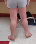
  •  Knock-knees or Flat foot
  •   Unusual or improper shaped patella under surface
  •   Arthritis

What are the signs and symptoms associated with Chondromalacia Patellae?

  • Chondromalacia Patellae generally leads to a vague discomfort in the front of the knee, generally known as patellofemoral pain. This pain worsens with the activities like; sitting, bending, standing for longer periods and exercising
  • Feeling of tightness in the knee area
  •  Reduction in thigh muscle mass
  • Minor swelling in the knee area
  • Loss of thigh muscle strength
  • Grinding or cracking sensation when knee is extended
  • Pain increases with kneeling down and squatting
  • You may feel trouble moving your knee joint past a certain point

What are the treatment options available to treat Chondromalacia Patellae?

Non-surgical treatment techniques are generally adopted to treat Chondromalacia Patellae. The most effective treatment technique is to adhere to a well-organised Physical Therapy treatment program. A Physical Therapy treatment program comprises of a complete rehabilitation program improving your muscle function and flexibility while relieving you from pain and swelling. Following treatment techniques are adopted under a complete Physical Therapy Rehabilitation Program:

Initially for the management of pain and swelling, physical therapists may recommend:

  • Resting your knee
  • Avoiding activities that irritate your knee cap
  • Icing your knee
  • Knee braces or arch supports to protect your knee joint  and to improve the alignment as well
  • Special shoes inserts and support devices (orthotics) for flat feet

Once the pain and swelling subsides below mentioned treatment techniques are employed by the Physical Therapists to help the patients to regain pain-free functioning and activity:

  • Selective strengthening exercises are recommended to strengthen the inner portion of thigh muscles
  • Stationary bicycling, pool running or swimming are advised to maintain cardiovascular conditioning
  •  Isometric exercises involving the tightening and releasing of muscles are recommended to maintain muscle mass
  • Taping of knee is recommended to reduce pain and as well to enhance the exercising ability
  • Specific exercises to correct the misalignment and muscle imbalance are recommended

Contact Alliance Physical Therapy

Contact Alliance Physical Therapy for the efficient and state-of-art treatment and quick relieve from any of your musculoskeletal pains. Our compassionate physical therapists make use of minimum invasive diagnostic and treatment techniques to save you from further pain and suffering and assist you to lead an active and healthy life. 

Skier’s Thumb: Causes, Symptoms and Treatments

What do we mean by Skier’s Thumb?

Skier’s Thumb is described as an injury caused to Ulnar Collateral Ligament (UCL) of thumb joint. The twisting and tearing away of UCL results this condition. Any injury to UCL weakens thumb’s gripping strength and as well causes loss of function. In other words, it unstables or loosens the thumb joint.

As this injury recurrently happens during skiing accidents, therefore it is referred to as Skier’s Thumb. But it does not imply that this injury occurs to skiers’ only. It can affect anybody who falls on an outstretched hand with sufficient force applied to thumb that extends it away from the index finger.

Skier's Thumb Treatment & Management

What are the causes of Skier’s Thumb?

Following conditions can be cited as the common causes of Skier’s Thumb:

  • A straight away or direct injury to your thumb
  • Extending away of thumb from the palm of your hand
  • Falling on your open hand with tucked in thumb
  • Repetitive and gradual traumas to your thumb
  • Slipping on the ski-slopes while holding or hands strapped to ski pole

What are the signs and symptoms of Skier’s Thumb?

  • Pain, tenderness and swelling at the inner base of your thumb
  • Bruising
  • Difficulty in holding objects and moving your thumb
  • Difficulty in applying force using your thumb
  • Ability to pinch and grasp things with thumb and index finger gets impaired
  • Difficulty in throwing objects
  • Unstable Thumb
  • A lump in your thumb
  • Deformed thumb

What are the treatment options available for Skier’s Thumb?

The treatment for Skier’s Thumb depends upon the extent of injury, patient’s condition and his specific needs and requirements. Following treatment options are followed to treat Skier’s Thumb:

  • To ease pain and swelling, ice pack may be placed on your thumb for 2 or 3 days after injury
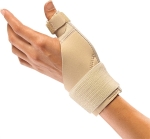
  • To immobilize or reduce the movement of thumb, support devices like; splint, brace or thumb cast may be used.

The splints or thumb casts are worn for 4 to 6 weeks and thereafter, a physical therapy regime is followed and adopted to mobilize the thumb again. It includes:

  • Ultra sound therapy; where sound waves are used to treat injury to soft tissues.
  • Massage Therapy is administered to repair the affected ligament.
  • Mobilization exercises are performed to restore thumb functioning and movement.
  • Physical Therapists make use of Hand Therapy balls and Therapeutic Putty to restore thumb strength and dexterity.
  • Strengthening exercises are performed to strengthen the otherwise loosened thumb.

Contact Alliance Physical Therapy for efficient treatment of any of your musculoskeletal problems. Our patients’ well being and care are our top most priorities. Our brigade of exuberant, determined and acknowledged physical therapists strives to provide finest clinical treatments to our patients in friendly and caring environment.

Identifying and Treating Cluster Headaches

What are Cluster Headaches?

Cluster Headaches are one-sided headaches which occur in cyclic patterns or clusters and are marked by the tearing of the eyes and stuffy nose. It is amongst the most agonizing and traumatic types of pain. Such headaches occur at the same time every year and is much more painful if it strikes you at night as compared to the day time. Episodes of pain occur constantly for one week to one year.

Who normally get affected by Cluster Headaches?

Men are at higher risk of getting affected by Cluster Headaches than women. These headaches can strike at any age but are more likely to occur at adolescent or middle age. These are commonly inherent and are transmitted through genes.

What triggers Cluster Headaches?

The exact causes of Cluster Headaches are still not known to the physicians, but  the following are considered as the potential causes of Cluster Headaches:

  • Sudden release of Histamine or Serotonin
  • In taking alcohol or Smoking cigarettes
  • Bright Lights
  • Exertion
  • Heat
  • Nitrites rich food
  • Cocaine
  • Stress and Anxiety
  • Season(common in spring or autumn)

What are the symptoms associated with Cluster Headaches?

These headaches arise frequently and without any warning signals but the pain most commonly attacks 2 or 3 hrs. after you fall asleep. Following can be enlisted as the signs and symptoms of Cluster Headaches:

  • Excessive or throbbing pain; commonly concentrated around or in one eye
  • One sided pain; from neck to temples
  • Uneasiness
  • Excessive Tearing
  • Redness in the affected side eye
  • One sided runny nose
  • Sweating
  • Swelling around the affected side eye
  • Drooping eyelids

What are the treatments available for Cluster Headaches?

Physicians adopt two types of techniques to treat Cluster Headaches viz.

  • Abortive Medications: These medications are prescribed to treat pain when it strikes. Imitrex or some other triptans are prescribed to subside pain when it occurs. At times, oxygen therapy is also administered, where you have to inhale oxygen through face mask.
  • Preventive Medications :  These medicines are prescribed by your doctor to minimize the time duration of cluster headaches and to reduce the severity of pain during cluster headaches.

How Physical Therapists can help to treat Cluster Headaches?

Physical Therapists can assist you in getting rid of  your Cluster Headaches beyond medications. Practicing exercises under the surveillance of an expert and trained Physical Therapist can enable you to relax muscle tension and as well to relieve stress. Physical Therapists prescribe following exercises to treat Cluster Headaches :

  •    Physical Therapists administer various relaxation techniques to help you to relax the muscles of your neck, jaw and shoulders. Progressive Relaxation techniques  are also administered and practiced to relax each and every muscle of your body.
  •    Cluster Headaches occur due to stress. Your breathing patterns get disturbed, when you are stressed. So to alter these disrupted breathing patterns, Physical Therapists practice different breathing exercises and make you relieved. These exercises are really advantageous when you are suffering from pain.
  •    Exercises that help to alleviate stress are followed. These help to reduce the occurrence ands severity of headaches. These also heighten the level of beta- endorphins which are your natural  stress relievers.

Contact Alliance Physical Therapy for the efficient management of any type of pain. Timely diagnosis and treatment can save you from further pain and suffering and as well help you to lead a healthy and quality life. 

Vestibular Rehabilitation Therapy

What Is Vestibular Rehabilitation Therapy?

Vestibular Rehabilitation Therapy (VRT) is a specific exercise based program that is designed and developed by a professional Physical Therapist to maximize the natural bodily capacities to compensate for the balance disorders and as well to lessen the dizziness related problems. Through Vestibular Rehabilitation Therapy physical therapists treat variety of symptoms related to vestibular disorders viz.

  • Dizziness
  • Vertigo
  • Imbalance
  • Nausea
  • Anxiety
  • Fatigue
  • Trouble concentrating
  • Blurred Vision
  • Muscle or Neck Stiffness or pain
  • Headaches

Which are the tests conducted under Vestibular Rehabilitation Therapy?

Before designing an efficient Vestibular Rehabilitation Therapy plan for a patient, following tests are conducted to recognize his symptoms and to identify problem areas:

  • Examination of patient’s medical history
  • Identifying and measuring balance, gait and posture
  • Eye- head Co-ordination tests
  • Questions are asked in form of Questionnaire to measure the severity of symptoms

What type of exercises is prescribed under Vestibular Rehabilitation Therapy?

The brain of a patient suffering from vestibular disorders can not depend upon the information it receives from vestibular system. As a result of this, his ability to maintain balance gets affected and the symptoms start getting worse and heightened, day by day. To overcome such conditions, vestibular exercises including the following are practiced under Vestibular Rehabilitation Therapy to improvise gait and gaze stability of a patient:

  • VOR Stimulation Exercises: This include the exercises which help to stabilize gaze and visual focus as well.During the course, the patient is asked to fix his vision on a set target while moving his or her head.
  • Ocular Motor Exercises:  This include a course of exercises, where the patient traces the field with the movement of his eyes only, i.e. without stirring his or her head.
  • Balance Retraining Exercises: This incorporates the set of exercises, which aid to better the co-ordination of muscular responses and structuring of sensory information as well, which in turn results in improved balance control.
  • Canal Re-positioning Maneuvers: These are administered to dislocate the debris with in the affected canal. This debris consists of small crystals of calcium carbonate that get collected with in the canal of inner ear.
  • Aquatic Physiotherapy: It is beneficial for the patients suffering from the problem of chronic dizziness. During the course, all of the exercises are administered in a pool.

 If the patients suffering from vestibular disorders perform their exercises correctly and regularly, in most of the cases the balance problems are improvised ad more over symptoms like muscle tension, dizziness, vertigo, headaches, fatigue begin to diminish or disappear completely with time.

Alliance Physical Therapy offers a wide range of support services to the patients. Our trained and qualified physical therapists provide reliable and customized treatment plans as per requirement and condition of the patients and help them to rehabilitate as soon as possible.

Physical Therapy Post Knee Replacement

Knee tends to be the most common joint to be replaced in the human body. People are advised Knee Replacement surgery if, either arthritis, bone diseases and some fracture has deteriorated the joint or if alignment problems have started hindering daily activities like walking, sitting and standing.

Post Knee Replacement Physical Therapy

Physical Therapy techniques after Knee Replacement Surgery help you to heal and resume your routine activities faster. Physical Therapist provides you with rehabilitation services during and after hospitalization.
While you are in the hospital after surgery, a physical therapist will :
•Educate you about practice walking with help of a walker or crutches
•Guide you regarding getting in and out of a chair or bed safely
•May employ electrical stimulation to decrease pain and swelling
•Administer isometric exercises to prevent blood clotting and to enhance blood circulation.
After you are discharged, your physical therapist will execute a customized treatment plan including various progressive exercises catering to your specific needs and goals as well :

•Motion Exercises : Pain and swelling can impede the progress of your replaced joint. Physical Therapists, to regain your motion, make you learn exercises that will enhance blood circulation and reduce swelling and as well prevent blood clotting. Below listed exercises may be performed to heighten the range of motion:
1.Ankle Pumps
2.Knee Bending
3.Heel Slides
4.Knee Straightening Stretch

•Strengthening Exercises : These exercises strengthen the otherwise weakened muscles of thigh and lower leg. Physical Therapists administer these exercises to strengthen your muscles and to stabilize knee joint. Following exercises are performed to revitalize your muscles :
1.Lying Kicks
2.Quadriceps Sets
3.Straight Leg Lifts
4. Exercycling
Balance Training: Specific balance training exercises are taught and performed to help patients to gain gait stability. These exercises are administered when you become able to put full weight and pressure on your replaced knee. These may include :
1.Leg Slides
2.Sitting Unsupported Knee Bends
3.Knee Exercises with Resistance
•Functional Training: Physical Therapists forward this training when you become capable to walk freely without feeling any pain or strain. This program is executed to heighten your activity level. They may include following exercises in your schedule :
1.Stair Climbing and Descending
2.Prolonged Knee Stretch
3.Standing Knee Bends
4.Community based actions viz. Crossing a busy street, Getting on and off escalator

Contact Alliance Physical Therapy for your successful recovery after Knee Replacement and to get back to your feet as soon as possible. We develop a customized treatment plan after collaborating with your consulting surgeon and will help you to rehabilitate quickly.

How to cure Plantar Fasciitis?

Symptoms and Treatment of Plantar Fasciitis:

images  3

Heel pain is a common complaint that has many causes. Heel pain may result from inflammation of the tissue on the bottom of the foot. This is called plantar fasciitis.

The plantar (foot) fascia (connective tissue) stretches under the skin across the arch of the foot from the heel to the base of the toes, when this tissue is tom, overused, or improperly stretched. It can become inflamed (fasciitis) Soreness, tenderness, and palm result. Persons who are overweight, female, or older than 40 years or who spend long hours on their feet are especially at risk of developing plantar fasciitis  Athletes, especially joggers and runners, may develop plantar fasciitis.

Sometimes plantar fasciitis can be associated with heel spurs. These spurs are outgrowths of bone on the calcaneus (heel bone). They are sometimes painful and may occasionally require surgical treatment.

Symptoms

  • Heel Pain, especially in the early morning or after a period of rest
  • Increasing pain with standing
  • Pain in the heel after exercising

Treatments

  • Rest
  • Arch supports (sometimes called orthotics) to be worn in shoes
  • Stretching the calf muscles and Achilles tendon
  • Ice packs
  • Nonsteroidal anti-inflammatory drugs (NSAIDs) such as ibuprofen or naproxen
  • Reducing excess body weight
  • Corticosteroid injections may be used in select cases
  • Surgery may be helpful if other treatments are not successful

Other causes of Heel Pain

It is important to understand that all heel pain is not from plantar fasciitis. Other medical problems can cause foot and heel pain. Diabetes and blood vessel disease, both serious medical problems, can cause heel pain. Arthritis, traumatic injury and bruising, gout, stress fractures (caused by repeated stress on bone), and other diseases can also cause heel pain. Rarely, tumors (either benign or cancerous) or infections can cause heel pain. If you develop persisting heel pain, see your doctor for an evaluation.

Plantar fasciitis is common in middle-aged people. It also happens in young individuals who are on their legs a lot, like sportsmen or military. It can occur in one feet or both legs. For best treatment of Plantar fasciitis Visit http://www.alliancephysicaltherapyva.com Contact Us at: 703-751-1008.