Tag Archives: Physical Therapy

How to cure Foot Pain?

Causes and Symptoms of Foot Pain:

Foot Pain

Foot pain can affect any part of your foot, from your toes to your Achilles tendon at the back of your heel. Some foot pain is just an annoyance, but foot pain can also be more serious, especially if it’s the result of an injury or certain chronic conditions. Minor foot pain usually responds well to home treatment but more severe foot pain needs medical attention. If not treated, some types of foot pain can lead to long-term damage or disability.

Causes of Foot Pain

Most foot pain is due to poorly fitting shoes, injury or overuse. But structural defects and medical conditions, such as arthritis and diabetes, also can lead to foot problems. Foot pain may be caused by many different conditions or injuries. Acute or repeated injury, disease, or a combination is the most common causes of foot pain. Injury is a result of forces outside the body either directly impacting the body or forcing the body into a position where a single or combination of forces results in damage to the structures of the body. Poor biomechanical alignment may lead to foot pain. Wearing shoes that are too tight or high heels can cause pain at the balls of the feet and the bones in that area. Shoes that are tied too tightly may cause pain and bruising on the top of the foot.

  • Injuries such as ligament sprains, bruises, muscle strains and fractures commonly happen suddenly (acutely).
  • Sprains, strains, bruises, and fractures may be the result of a single or a combination of stresses to the foot.
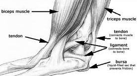
  • A sprain of the foot or ankle happens when ligaments that hold the bones together are overstretched and their fibers tear.
  • The looseness of ligaments in the joints of the foot may lead to foot pain.

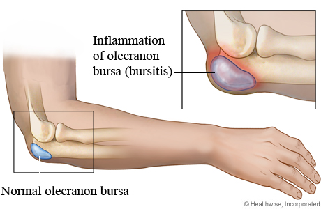
The muscle’s bursa and fascia of the foot can be strained by overstretching, overuse, overloading, bruising, or a cut (such as by stepping on a sharp object). Achilles tendonitis is a common injury to the tendon that attaches at the back of the heel.

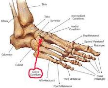
  • Injury to the bones and joints of the foot can be caused by a single blow or twist to the foot, or also by repetitive injury that can result in a stress fracture.
  • A blunt-force injury such as someone stepping on your foot may result not only in a bruise (contusion) injury but also damage to the muscles and ligaments of the foot.
  • Direct blows to the foot can cause bruising, breaking of the skin, or even fracturing of bones.
  • Metarsalgia is the irritation of the joints of the ball of the foot. “Turf toe” is a common athletic injury in which the tendon under the joint at the base of the big toe is strained.
  • Injury to the toenail can cause pooling of blood under the nail and the permanent or temporary loss of a toenail.
  • Repetitive injury to the bones, muscles, and ligaments can result in extra bone growth known as spurs or exostosis.

Symptoms may accompany foot pain

Pain and point tenderness are the first indications that something is wrong in a specific area. The onset of pain, whether suddenly or over time, is an important indicator of the cause of the problem.

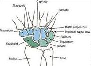
Bones of the foot are joined together by ligaments. A sprain happens when the ligaments that hold the bones together are overstretched and the fibers tear. Point tenderness and looseness of a joint are indications of a sprain.

Injury to the bones of the foot can be caused by a single blow or twist to the arch or also by repetitive injury that can end in a stress fracture. Fractures are indicated by a small point of pain that may be exquisitely tender on the bone. There may be a noticeable lump or gap at the site of the fracture. A turned toe or forefoot may also be a sign of a fracture.

Injury to the bones of the foot can be caused by a single blow or twist to the arch or also by repetitive injury that can end in a stress fracture. Fractures are indicated by a small point of pain that may be exquisitely tender on the bone. There may be a noticeable lump or gap at the site of the fracture. A turned toe or forefoot may also be a sign of a fracture.

Prevention of Foot Pain

  • To prevent injuries and pain, the following issues should be addressed before starting an exercise routine.
  • Are you in good health? A general physical exam by a physician will help to evaluate your cardiovascular function, the possibility of disease or any other general medical problems that you may have.
  • Before beginning activities, diseases such as gout, diabetes, certain types of arthritis, and neuropathies should be treated.

Treatment for Foot Pain

When the pain begins to interfere with your daily living activities or if you cannot perform your chosen activities without pain, you should consider getting medical attention. Indications that you should seek medical care are:

The area looks deformed, you have loss of function, change of sensation, a large amount of swelling with pain, prolonged change of skin or toenail color, the affected area becomes warmer than the surrounding areas, becomes extremely tender to the touch, or is causing you to move differently.

At Alliance Physical Therapy we provide 24/7 access to online appointments, with most of the requests scheduled in less than 48 hours. Visit here for more information: http://www.alliancephysicaltherapyva.com

What are the Causes of Achilles Tendonitis (Heel Pain)?

Symptoms and Causes of Achilles Tendonitis:

Achilles Tendonitis is inflammation of the Achilles tendon. In most cases, it is a type of overuse injury and is more common in younger people. Professional and weekend athletes can suffer from Achilles tendonitis, but it is also a common overuse injury in people not involved in sport. Treatment includes rest, non-steroidal anti-inflammatory drugs (NSAIDs), physical therapy and avoiding activities that aggravate the condition.

Achilles Tendonitis

Symptoms of Achilles Tendonitis

Symptoms of Achilles Tendonitis include:

  • Pain in the back of the heel
  • Difficulty walking – sometimes the pain makes walking impossible
  • Swellingtenderness and warmth of the Achilles tendon

Causes of Achilles Tendonitis

Some of the causes of Achilles tendonitis include:

  • Overuse injury –This occurs when the Achilles tendon is stressed until it develops small tears. Runners seem to be the most susceptible. People who play sports that involve jumping, such as basketball, are also at increased risk.
  • Arthritis – Achilles tendonitis can be a part of generalised inflammatory arthritis, such as ankylosing spondylitis or psoriatic arthritis. In these conditions both tendons can be affected.
  • Foot problems – Some people with flat feet or hyperpronated feet (feet that turn inward while walking) are prone to Achilles tendonitis. The flattened arch pulls on calf muscles and keeps the Achilles tendon under tight strain. This constant mechanical stress on the heel and tendon can cause inflammation, pain and swelling of the tendon. Being overweight can make the problem worse.
  • Footwear – Wearing shoes with minimal support while walking or running can increase the risk, as can wearing high heels.
  • Overweight and obesity – Being overweight places more strain on many parts of the body, including the Achilles tendon.

Diagnosis of Achilles Tendonitis

To confirm the diagnosis and consider what might be causing the problem, it’s important to see your doctor or a physiotherapist. Methods used to make a diagnosis may include:

  • Medical history, including your exercise habits and footwear.
  • Physical examination, especially examining for thickness and tenderness of the Achilles tendon.
  • Tests may be required. These may include an x-ray of the foot, ultrasound and occasionally blood tests (to test for an inflammatory condition), and an MRI scan of the tendon.

Treatment for Achilles Tendonitis

The aim of the treatment is to reduce strain on the tendon and reduce inflammation. Strain may be reduced by:

  1. Avoiding or severely limiting activities that may aggravate the condition, such as running.
  2. Using shoe inserts (orthoses) to take pressure off the tendon as it heals. In cases of flat or hyperpronated feet, your doctor or podiatrist may recommend long-term use of orthoses.
  3. Inflammation may be reduced by:
  • Applying icepacks for 20 minutes per hour during the acute stage
  • Taking non-steroidal anti-inflammatory drugs

Other Treatment that may be Recommended

You may also be given specific exercises to gently stretch the calf muscles once the acute stage of inflammation has settled down. Your doctor or physiotherapist will recommend these exercises when you are on the road to recovery. Recovery is often slow and will depend on the severity of the condition and how carefully you follow the treatment and care instructions you are given.

Alliance  Physical Therapy is the Best Physical Therapy Clinic. We provide 24/7 access to online appointments, with most of the requests scheduled in less than 48 hours. Call now for Quick Appointment: 703-356-3470

http://www.alliancephysicaltherapyva.com

How to get Relaxation from Neck Pain?

Causes of Neck Pain:

index

Neck pain can be caused by irregularities in the soft tissues areas, namely in the muscles, ligaments, and nerves. The irregularities can also occur in the bones and joints of the spine. The most common causes of neck pain in the soft-tissue regions are due to injuries, degeneration, or prolonged wear and tear on the muscles and ligaments. In rare cases, infections or tumors may be the cause of neck pain. For some people, neck problems may be the source of pain in the upper back, shoulders, or arms.

Other causes of neck pain can be caused by:

INFLAMMATORY DISEASES - Rheumatoid arthritis can cause destruction of the joints of the neck. Rheumatoid arthritis typically occurs in the upper neck area.

CERVICAL DISK DEGENERATION - The cervical discs act as a shock absorber between the bones in the neck. In cervical disk degeneration, which typically occurs in people age 40 years and older, the normal gelatin-like center of the disk degenerates and the space between the vertebrae narrows. As the disc space becomes narrow, additional stress is placed on the joints of the spine.

INJURY - Because the neck has so much flexibility and it must constantly support the head, it is extremely susceptible to injury. Motor vehicle or diving accidents, participation in contact sports, traumatic injuries, or falls may result in neck injuries. The regular use of safety belts in motor vehicles can help to minimize or prevent neck injury.

OTHER CAUSES - Tumors, Infections, or Congenital abnormalities of the vertebrae may also cause neck pain and range of motion limitations.

Risks and Prevention of Neck Pain

You may be most shocked to learn that you do much of your standing, sitting, exercise and other activities with a forward head. Test yourself and see if you have a tendency to tilt forward:

  • Stand with your back to a wall, but not touching the wall.
  • Back yourself up to the wall until something on your body makes contact.

Start paying attention to how other people sit while eating or how they tend to carry a large purse or backpack. Does their neck tilt forward against the load or are they using muscles to hold the spine in a healthy position? The average person will overstretch their neck and upper body unequally so often, it is a mystery that they don’t have more pain.

Cure-Your-Neck-Pain-Ebook

  • Stand with your whole body (heels, hips, upper back, and the back of your head) against a wall.
  • Bring the back of your head against the wall without raising or dropping your chin, or arching your back.
  • If you cannot keep your heels, hips, upper back, and the back of your head against the wall in a comfortable position or you crane your neck, you are too tight to stand up straight.

Many people are susceptible to neck and shoulder pain because of repetitive work-related issues, poor posture, and overall bad habits. The things listed below will help you assess your situation and your likely cause of neck and shoulder pain.

Be aware of your Posture

If you are sitting in the same position for long amounts of time you are a prime canditate for neck stiffness and pain.

  • Identify the risks associated with your job and your daily work routine. If you are required to lift heavy objects or are at risk of injury due to a fall or other trauma, you may eventually sustain a more serious injury which causes neck pain. Be certain you take the necessary precautions and safety measures while working.
  • Make an appointment with your doctor for a full check-up annually, especially if you are at increased risk of osteoporosis or congenital problems.
  • Examine your lifestyle for habits that may lead to pain. Pay attention to how you fall asleep, or when you are resting on the couch. Pillows that are very soft or filled with feather or down are likely to cause bad posture during sleep. You should investigate pillows that support the natural curve of your back and neck. Make sure you have suitable furniture in your home. A desk chair that is not supportive or a pillow that doesn’t offer enough neck support will lead to bad posture and result in neck pain.
  • Use relaxation techniques when you are under mental and emotional stress. Stress is a major culprit in bad cases of neck pain. Incorporate the following daily exercises and activities into your day to keep muscles flexible and healthy. Exercising regularly will also reduce tension and stress hormones in your body.

Aquatic therapy or pool therapy consists of an exercise program that is executed in the water. It is a valuable form of therapy that is useful for a range of medical conditions. Aquatic therapy utilizes the physical properties of water to aid in patient healing and exercise performance. Call today at: 703-670-9935

http://www.alliancephysicaltherapyva.com

Mechanisms of Spinal Cord Injury

Causes and Syndromes of Spinal Cord Injury:

images

Injuries to the cervical spine constitute uncommon but nonetheless devastating occurrences to those participating in athletic events. These injuries happen primarily to athletes involved in the contact sports of football, wrestling, and ice hockey, with football injuries constituting the largest number of cases. The important work of Schneider in the mid 1960s, focusing on football-related head and neck injuries, resulted in a significant reduction in the incidence of these accidents owing to improvements in equipment, education in proper techniques, offseason conditioning, and rule changes.

Causes and Mechanisms of Spinal Cord Injury

  • The mechanism of spinal cord injury can be sport related, but it is more commonly independent of the sport.
  • The major mechanism of serious cervical injury is an axial load, or a large compressive force applied to the top of the head.
  • This mechanism is more dangerous when the neck is slightly flexed, because the spine is brought out of its normal lordotic alignment, which does not allow for proper distribution of force to the thorax.
  • Flexion puts the cervical spine in a straight line; thus, the musculature cannot assist in absorbing the force.
  • Torget al indicated that injury occurs to the cervical spine when it is compressed between the body and the rapidly decelerating head.
  • When a fracture results, if the bone fragments or herniated disc materials encroach on the spinal cord, neurologic damage will occur.
  • This mechanism is the primary cause of cervical fracture, dislocation, and quadriplegia.

Syndromes of Spinal Cord Injury

Spinal trauma may result in a variety of clinical syndromes, according to the type and severity of the impact and bony displacement as well as the subsequent secondary insults such as:

Complete spinal cord injury is a transverse myelopathy with total loss of spinal function below the level of the lesion. This insult is caused by either anatomic disruption of the spinal cord or, more commonly, hemorrhagic or ischemic damage at the site of injury. Although the spinal cord usually remains in continuity, a physiologic block to impulse transmission results in the complete injury. Complete injury patterns are rarely reversible, although with long-term follow-up, improvement of 1 spinal level may be seen when the initial segmental traumatic spinal cord swelling resolves.

  • Several patterns of incomplete spinal cord injury are common, usually produced on a vascular basis.
  • The central cord syndrome causes incomplete loss of motor function with a disproportionate weakness of the upper extremities as compared with the lower extremities.
  • This condition is thought to be the result of hemorrhagic and ischemic injury to the corticospinal tracts because of their somatotopic arrangement.

Fibers of cervical nerves that innervate the upper extremities are arranged more medially than those subserving function to the lower extremities. The originally described central cord syndrome also includes nonspecific sensory loss and bladder and sexual dysfunction. This injury pattern is also often seen in older patients with vertebral osteophytes and in those with hyper-extension injuries.

Alliance Physical Therapy provide a comprehensive, multi-disciplinary approach to neurological rehabilitation that includes muscle and sensory re-education, coordination activities, range of motion and speech therapy to those patients who have experienced a neurological disorder such as a stroke or spinal cord injury. Call now for Quick Appointment: 703-205-1919

http://www.alliancephysicaltherapyva.com

Physical Therapy treatment for Back Pain

Treatment for Back Pain:

index

Back pain and injuries can be treated and managed extremely well using a range of physical therapies in combination with controlled exercise programmes. Often this is combined with medication to provide patients with a good outcome and relief of their pain and symptoms. The aim of this approach is to:

  • Decrease pain and stiffness
  • Improve daily quality of life
  • Improve and maintain fitness, strength and posture
  • Increase flexibility and endurance

Several avenues and professionals people are available to help with this conservative treatment.

Exercise

  • People who are physically fit usually suffer less back pain, and recover from injuries more quickly.
  • The muscular supports of the spine are crucial to maintaining good function and stability of the back.
  • With ageing, injury or underlying spinal problems, the back muscles can easily become weakened, lose tone and are quickly tired.
  • It’s known that poor muscle condition can in itself be a cause of disability and pain.
  • Bed rest for more than one or two days can delay recovery and slow progress, as it results in joint mobility loss and deterioration in physical fitness.
  • A programme should include specific exercises to help your condition as well as to strengthen your back, leg and abdominal muscles.
  • If you have pain during the exercise, it may mean you’re not doing it correctly or that it’s not suitable for you. Talk to your doctor or physiotherapist.
  • Walking, swimming and riding an exercise bike are good ways to improve fitness, but always seek professional advice, as some may aggravate back problem (for example, some swimming strokes).

Physical Therapy

Using a variety of techniques and equipment (including Pilates), and working closely with the patient, a physiotherapist is professionally trained to:

  • Assess and treat your back pain with medical consultation and in partnership with your spinal surgeon
  • Help you restore normal back and spinal function
  • Help you learn how to prevent further injury
  • Provide specific exercises for your back condition

Our Lower Back Program uses a standardized, functional and outcomes-oriented approach to care that focuses specifically on the lower back. Through an active and educational continuum of treatment, our therapists assist the patient in returning to normal, daily activities as soon as possible. By providing comprehensive education in anatomy, pathology and care of the lower back, the patient is empowered to participate in his or her recovery and in the prevention of future injury. Call now for Best Physical Therapy: 703-704-5771

http://www.alliancephysicaltherapyva.com

How to cure Wrist Bone Fracture?

Classification and Treatment of Wrist Bone Fracture:

images 1

Trapezium bone accidents are unusual carpal bone accidents. They can either happen in solitude or along with other carpal bony injury. This accounts for 1 to 5 percent of wrist fractures. It could be isolated fracture or dislocations.

Mechanism of Injury

  • Fall on an outstretched hand.
  • Direct blow over the dorsum of the hand.

Classifications

Trapezium fractures are divided into:

  • Body fractures
  • Ridge fractures (Palmar)
  • Dislocations: This could be dorsal, palmar or radial and may be associated with fracture of the scaphoid and trapezium.

Clinical Features

The patient complains of:

Investigations

Plain X-rays though useful are not reliable. CT scan is a better option.

Treatment

  • Undisplaced fracture: Thumb spica for 4 to 6 weeks.
  • Displaced fracture: Open reduction and rigid internal fixation is advised.
  • Dislocation is treated by open reduction and K-wire fixation.

Alliance Hand Therapy is currently providing care throughout Northern Virginia from our clinics located in Alexandria, Fairfax, Springfield and Woodbridge. Our Hand therapy Program is a specialized treatment program focusing primarily on conditions affecting the hand and upper extremities. Call now at: 703-750-1204 or Visit: http://www.alliancephysicaltherapyva.com

What are the classifications of Capitellum (Elbow) Fracture?

Classifications and Treatment of Capitellum Fracture (Elbow Injury):

images

Capitellum is the anterior portion of the lateral humeral condyle. This fracture is unique in being intra-articular always.

Mechanism of injury

Fall on an outstretched hand, with flexion or extension of the elbow and the resulting shear forces through the radial head slices the capitellum.

Classifications

Based on the size of the articulating fragment, it is classified into three types:

  • Type I (Hahn-Steinthal variety): This involves a large portion of the capitellum and a small chunk of trochlea with less of subchondral portion.
  • Type II (Kocher-Lorenz variety): Here only a large portion of the capitellum is involved with a huge chunk of subchondral bone.
  • Type III: Comminuted fracture.

Clinical Features

  • The patient complains of pain and swelling over the lateral aspect of the elbow.
  • Elbow and forearm movements are also restricted.

Radiographs

A true lateral view of the elbow is mandatory to accurately diagnose this fracture. The characteristic finding of this fracture is the presence of “double arc sign” described by McKay over the X-ray.

Treatment

  • Undisplaced fractures can be managed conservatively by an above elbow plaster cast or slab for 3 to 4 weeks.
  • Displaced fractures need open reduction and internal fixation with minifragment screws.

Alliance Hand Therapy is currently providing care throughout Northern Virginia from our clinics located in Alexandria, Fairfax, Springfield and Woodbridge. Our Hand therapy Program is a specialized treatment program focusing primarily on conditions affecting the hand and upper extremities. Our Certified Hand Therapists have a high degree of specialization that requires several thousands of hours continuing education and advanced certification. Call today for best Hand Therapy: 703-726-9352

http://www.alliancephysicaltherapyva.com

How to prevent Foot Bone Injury?

Clinical Features and Investigation of Foot Bone Injury:

images

Foot injuries are rare injuries and are usually due to indirect forces. More commonly, they are associated with injuries to the tarsometatarsal joints.

Clinical Features

Investigations

Plain X-ray (AP, lateral, oblique views) with CT scan of the foot.

Classification

Group A:  Extra-articular

Group B:  Partly intra-articular (involves other navicular cuneiform or metatarsal cuneiform joints).

Group C:  Involves both articular surfaces.

Treatment

Non-operative: Short leg cast for 6 to 8 weeks for undisplaced fractures.

Operative: For displaced fractures, open reduction and internal fixation with pins or screws.

Alliance Physical Therapy provide 24/7 access to online appointments, with most of the requests scheduled in less than 48 hours. If you are suffering from Foot Bone Injury then visit http://www.alliancephysicaltherapyva.com/ or contact us at: 703-670-9935.

How to get relief from Wrist Joint Pain?

Clinical Features and Treatment of Wrist Joint Injury:

images 1

Wrist Joint is a common carpal dislocation and can lead to severe disability of the wrist function.

Mechanism of injury

This is due usually due to fall on the out-stretched hands. It can cause late carpal instability and arthritis. Hence, prompt and correct treatment is mandatory.

Clinical Features

  • Patient presents with pain
  • Swelling
  • Tenderness
  • Loss of wrist movements.

Radiograph

  • In radiograph of the lateral view, normally lunate forms a half-moon shape, which is lost in this dislocation.
  • Moreover, in the anteroposterior view the normal rectangular profile is lost.

Treatment

Problems

  • This may cause compression of the median nerve.
  • If left untreated it may cause permanent palsy, hence, reduction should be carried out as an emergency procedure.

Methods

  • If seen early, reduction is easy and immobilization for 3 weeks with wrist in slight flexion usually gives good results.
  • If seen after 3 weeks, open reduction is done.
  • If lunate cannot be reduced by open reduction, resection of the proximal carpal bones or arthrodesis of the wrist may be necessary.

Alliance Hand Therapy is currently providing care throughout Northern Virginia from our clinics located in Alexandria, Fairfax, Springfield and Woodbridge. Call today at: 703-205-1919

http://www.alliancephysicaltherapyva.com

How do you heal OLECRANON BURSITIS?

Elbow PainThis is a chronic inflammation of the olecranon bursa. It may be the result of repetitive minor injuries or irritation, microcrystalline deposition. Infection occurs due to chronic friction as in students who tend to keep their elbows repeatedly over the table, bench, etc. over long periods during writing, reading, etc.

Clinical Features:

It usually manifests as a swelling over the tip of the olecranon. There may be pain, if there is inflammation. Inspection or palpation usually easily detects it.

 Investigations:

Aspiration and culture of the bursal fluid are necessary in order to exclude the possibility of an infectious etiology.

 Treatment:

Treatment is essentially conservative and consists of NSAIDs, local steroids, etc. Surgical excision is done in chronic cases. Microcrystalline-induced bursitis has a good prognosis and the symptoms usually resolve after a few days, whether treated or not. However, bursitis due to repeated minor irritation is more difficult to treat.

Do not worry about Olercranon Bursitis now. We are here to diagnose you. Alliance Physical Therapy is best Rehab & Physical Therapy center in Virginia. Call now for quick Appointment: 703-704-5771

Visit our website now: http://www.alliancephysicaltherapyva.com