Tag Archives: Physical Therapy Clinic

How to cure Wrist Bone Fracture?

Classification and Treatment of Wrist Bone Fracture:

images 1

Trapezium bone accidents are unusual carpal bone accidents. They can either happen in solitude or along with other carpal bony injury. This accounts for 1 to 5 percent of wrist fractures. It could be isolated fracture or dislocations.

Mechanism of Injury

  • Fall on an outstretched hand.
  • Direct blow over the dorsum of the hand.

Classifications

Trapezium fractures are divided into:

  • Body fractures
  • Ridge fractures (Palmar)
  • Dislocations: This could be dorsal, palmar or radial and may be associated with fracture of the scaphoid and trapezium.

Clinical Features

The patient complains of:

Investigations

Plain X-rays though useful are not reliable. CT scan is a better option.

Treatment

  • Undisplaced fracture: Thumb spica for 4 to 6 weeks.
  • Displaced fracture: Open reduction and rigid internal fixation is advised.
  • Dislocation is treated by open reduction and K-wire fixation.

Alliance Hand Therapy is currently providing care throughout Northern Virginia from our clinics located in Alexandria, Fairfax, Springfield and Woodbridge. Our Hand therapy Program is a specialized treatment program focusing primarily on conditions affecting the hand and upper extremities. Call now at: 703-750-1204 or Visit: http://www.alliancephysicaltherapyva.com

What are the classifications of Capitellum (Elbow) Fracture?

Classifications and Treatment of Capitellum Fracture (Elbow Injury):

images

Capitellum is the anterior portion of the lateral humeral condyle. This fracture is unique in being intra-articular always.

Mechanism of injury

Fall on an outstretched hand, with flexion or extension of the elbow and the resulting shear forces through the radial head slices the capitellum.

Classifications

Based on the size of the articulating fragment, it is classified into three types:

  • Type I (Hahn-Steinthal variety): This involves a large portion of the capitellum and a small chunk of trochlea with less of subchondral portion.
  • Type II (Kocher-Lorenz variety): Here only a large portion of the capitellum is involved with a huge chunk of subchondral bone.
  • Type III: Comminuted fracture.

Clinical Features

  • The patient complains of pain and swelling over the lateral aspect of the elbow.
  • Elbow and forearm movements are also restricted.

Radiographs

A true lateral view of the elbow is mandatory to accurately diagnose this fracture. The characteristic finding of this fracture is the presence of “double arc sign” described by McKay over the X-ray.

Treatment

  • Undisplaced fractures can be managed conservatively by an above elbow plaster cast or slab for 3 to 4 weeks.
  • Displaced fractures need open reduction and internal fixation with minifragment screws.

Alliance Hand Therapy is currently providing care throughout Northern Virginia from our clinics located in Alexandria, Fairfax, Springfield and Woodbridge. Our Hand therapy Program is a specialized treatment program focusing primarily on conditions affecting the hand and upper extremities. Our Certified Hand Therapists have a high degree of specialization that requires several thousands of hours continuing education and advanced certification. Call today for best Hand Therapy: 703-726-9352

http://www.alliancephysicaltherapyva.com

How to prevent Foot Bone Injury?

Clinical Features and Investigation of Foot Bone Injury:

images

Foot injuries are rare injuries and are usually due to indirect forces. More commonly, they are associated with injuries to the tarsometatarsal joints.

Clinical Features

Investigations

Plain X-ray (AP, lateral, oblique views) with CT scan of the foot.

Classification

Group A:  Extra-articular

Group B:  Partly intra-articular (involves other navicular cuneiform or metatarsal cuneiform joints).

Group C:  Involves both articular surfaces.

Treatment

Non-operative: Short leg cast for 6 to 8 weeks for undisplaced fractures.

Operative: For displaced fractures, open reduction and internal fixation with pins or screws.

Alliance Physical Therapy provide 24/7 access to online appointments, with most of the requests scheduled in less than 48 hours. If you are suffering from Foot Bone Injury then visit http://www.alliancephysicaltherapyva.com/ or contact us at: 703-670-9935.

How to get relief from Wrist Joint Pain?

Clinical Features and Treatment of Wrist Joint Injury:

images 1

Wrist Joint is a common carpal dislocation and can lead to severe disability of the wrist function.

Mechanism of injury

This is due usually due to fall on the out-stretched hands. It can cause late carpal instability and arthritis. Hence, prompt and correct treatment is mandatory.

Clinical Features

  • Patient presents with pain
  • Swelling
  • Tenderness
  • Loss of wrist movements.

Radiograph

  • In radiograph of the lateral view, normally lunate forms a half-moon shape, which is lost in this dislocation.
  • Moreover, in the anteroposterior view the normal rectangular profile is lost.

Treatment

Problems

  • This may cause compression of the median nerve.
  • If left untreated it may cause permanent palsy, hence, reduction should be carried out as an emergency procedure.

Methods

  • If seen early, reduction is easy and immobilization for 3 weeks with wrist in slight flexion usually gives good results.
  • If seen after 3 weeks, open reduction is done.
  • If lunate cannot be reduced by open reduction, resection of the proximal carpal bones or arthrodesis of the wrist may be necessary.

Alliance Hand Therapy is currently providing care throughout Northern Virginia from our clinics located in Alexandria, Fairfax, Springfield and Woodbridge. Call today at: 703-205-1919

http://www.alliancephysicaltherapyva.com

How do you heal OLECRANON BURSITIS?

Elbow PainThis is a chronic inflammation of the olecranon bursa. It may be the result of repetitive minor injuries or irritation, microcrystalline deposition. Infection occurs due to chronic friction as in students who tend to keep their elbows repeatedly over the table, bench, etc. over long periods during writing, reading, etc.

Clinical Features:

It usually manifests as a swelling over the tip of the olecranon. There may be pain, if there is inflammation. Inspection or palpation usually easily detects it.

 Investigations:

Aspiration and culture of the bursal fluid are necessary in order to exclude the possibility of an infectious etiology.

 Treatment:

Treatment is essentially conservative and consists of NSAIDs, local steroids, etc. Surgical excision is done in chronic cases. Microcrystalline-induced bursitis has a good prognosis and the symptoms usually resolve after a few days, whether treated or not. However, bursitis due to repeated minor irritation is more difficult to treat.

Do not worry about Olercranon Bursitis now. We are here to diagnose you. Alliance Physical Therapy is best Rehab & Physical Therapy center in Virginia. Call now for quick Appointment: 703-704-5771

Visit our website now: http://www.alliancephysicaltherapyva.com

How to cure Ankle Tibial Nerve?

Clinical Features, Investigation and Treatment of Tibial Nerve:

images 22Stress fracture of the medial malleolus is an unusual injury but should be considered in the runner presenting with persistent medial ankle pain aggravated by activity. Although the fracture line is frequently vertical from the junction of the tibial plafond and the medial malleolus, it may arch obliquely from the junction to the distal tibial metaphysis.

Clinical Features

  • Athletes classically present with medial ankle pain that progressively increases with running and jumping activities.
  • Often they experience an acute episode, which leads to their seeking medical attention.
  • Examination reveals tenderness overlying the medial malleolus frequently in conjunction with an ankle effusion.

Investigations

  • In the early stages, X-rays may be normal, but with time a linear area of hyperlucency may be apparent, progressing to a lytic area and fracture line.
  • If the X-ray is normal, a radioisotopic bone scan, CT or MRI will be required to demonstrate the fracture.

Treatment

  • If no fracture or an undisplaced fracture is evident on X-ray, treatment requires weight-bearing rest with an air-cast brace until local tenderness resolves, a period of approximately six weeks.
  • If, however, a displaced fracture or a fracture that has progressed to non-union is present, surgery with internal fixation is required.
  • Following fracture healing, the practitioner should assess biomechanics and footwear. A graduated return to activity is required.

Stress bone injuries of the inside malleolus generally happen over time with extreme standing and walking action such as running. Physical rehabilitation treatment is essential for all sufferers with a stress crack of the inside malleolus to speed up treatment, avoid repeat and make sure an maximum result. At Alliance Physical Therapy we provide 24/7 access to online appointments, with most of the requests scheduled in less than 48 hours. For Best Rehabilitation and Physical Therapy Call now at: 703-750-1204

http://www.alliancephysicaltherapyva.com

How Foot and Ankle Discomfort is treated?

Causes and Treatment of Hallux Valgus:    Hallux valgus anatomy

Hallux Valgus is defined as a static subluxation of the first metatarsophalangeal joint. It is characterized by valgus (lateral) deviation of the great toe and varus (medial) deviation of the first metatarsal. Bony exostoses develop around the first metatarsophalangeal joint, often with an overlying bursitis. In severe cases, exostoses limit first metatarsophalangeal joint range of motion and cause pain with the pressure of footwear.

Causes

The development of hallux valgus appears to occur secondary to a combination of intrinsic and extrinsic causes. Recognized causative factors include:

  • Constricting footwear (e.g. high heels)
  • Excessive pronation-increased pressure on the medial border of the hallux, resulting in deformation of the medial capsular structures.
  • Others-cystic degeneration of the medial capsule, Achilles tendon contracture, neuro-muscular disorders, collagen deficient diseases.

Clinical Features

  • In the early phases hallux valgus is often asymptomatic, however, as the deformity develops, pain over the medial eminence occurs.
  • The pain is typicaIly relieved by removing the shoes or by wearing soft, flexible, wide-toed shoes. Blistering of the skin or development of an inflamed bursa over the medial eminence may occur.
  • In severe deformity, lateral metatarsalgia may occur due to the diminished weight-bearing capacity of the first ray.
  • Examination reveals the hallux valgus deformity often with a tender swelling overlying the medial eminence.

Investigation

Plain X-rays should be performed to assess both the severity of the deformity and the degree of first metatarsophalangeal joint degeneration.

Treatment

  • Initial treatment involves appropriate padding and footwear to reduce friction over the medial eminence.
  • Correction of foot function with orthoses is essential.
  • In more severe cases surgery may be required to reconstruct the first metatarsophalangeal joint and remove the bony exostoses.
  • Orthoses are often required after surgery.

Hallux valgus is a situation that impacts the combined at the platform of the big toe. This condition is commonly known as bunion. The big toe of the feet is known as the hallux. If the big toe begins to vary inward towards the child toe the situation is known as hallux valgus. Alliance Physical Therapy is best Rehab & Physical Therapy center in Virginia. Call now for quick Appointment: 703-704-5771

http://www.alliancephysicaltherapyva.com

How to get comfort from Thoracic Outlet Syndrome (Neck Tingling)?

Clinical Features and Treatment of Thoracic Outlet Syndrome:

Neck Tingling

The space at the thoracic outlet or inlet when it is less than adequate, subjects the neurovascular structures seeking to gain entry into the upper limbs via this space, to undue pressure. The blame for the neurovascular complaints should be placed at the doorstep of the decreased space and not at the structures producing the problems.

This syndrome results from the compression of neurovascular bundle comprising of subclavian artery and veinaxillary artery and vein and brachial plexus at the thoracic outlet. Thoracic outlet is a space between the first rib, clavicle, and the scalene muscles.

Sites of Compression

The sites of compression could be either Supraclavicular, Subclavicular or Infraclavicular.

  • Supraclavicular: Interscalene triangle between the anterior scalene muscles.
  • Subclavicular: Interval between the second thoracic rib, clavicle, and subclavius.
  • Infraclavicular: Beneath an enclosure formed by the coracoid process, pectoralis minor, and costocoracoid membrane.

Rare Cause

Scissor-like encirclement of axillary artery by the median nerve.

Contributing Factors

Dynamic Factors

Arm when in full abduction pulls up the artery by 180 degree causing compression in the short retroclavicular space.

Static Factors

  • Vigorous occupation: Increases the muscle bulk and thereby decreases the space.
  • Inactive occupation: Decreases the muscle bulk and thereby increases the space.
  • Congenital: Cervical rib decreases the interscalene space and thereby decreases the retroclavicular space.
  • Traumatic: Malunion or nonunion of fracture clavicle.

Anomalies of the first thoracic rib.

Miscellaneous

Clinical Features

Obviously, this syndrome poses two major problems. The first one relates to the compression of the major vessels and secondly to the compression of the nerves.

1.   Vascular Problems

Here the compression could be arterial or venous. During the arterial compression, which is mild in the early stages the patient complains of numbness of the whole arm with rapid fatigue during overhead exercises. If the compression is significant, the patient will complain of cold, cyanosis, pallor, and Raynaud’s phenomenon. Venous compression leaves the limb swollen and discolored after exercises, which disappears slowly with rest.

2.   Neurogenic Problems

Patients complain of par esthesia along the medial aspect of the arm, hand, little and ring fingers. There is weakness of the hand also.

Complications

  • Subclavian artery compression
  • Results in poststenotic dilatation
  • Stasis favors thrombosis
  • The thrombi break and migrate distally causing embolization

Investigations

  • Nerve Conduction Studies: Difficult to determine the nerve conduction velocity through the thoracic outlet, but its biggest value is to rule-out problems like entrapment, e.g. ulnar nerve at elbowwrist, etc.

Treatment

  • Conservative treatment: Consists of rest, physiotherapy, exercises like shoulder shrugging, etc.
  • Surgical treatment

Thoracic Outlet problem is a number of conditions that happen when the veins or anxiety in the thoracic store — the area between your collarbone and your first rib — become compacted. This can cause discomfort in shoulder area and throat and pins and needles in your fingertips. Call now for best Physical Therapy: 703-205-1919

http://www.alliancephysicaltherapyva.com

How we treat Fracture of Femur?

Classification And Treatment in Supracondylar Fracture of Femur:SUPRACONDYLAR FRACTURE OF FEMUR

Supracondylar region extends from the femoral condyles to the junction of metaphysis with femoral shaft .The distal fragment is displaced and angulated posteriorly due to the pull of gastrocnemius muscle.

Mechanism of Injury

It is due to severe valgus or varus forces with axial loading and rotation due to RTA, fall, etc.

Classification

  1. Nears’s Classification
  • Undisplaced Fracture
  • Displaced Fracture
  • Medial Displacement
  • Lateral Displacement
  • Comminuted Fracture
  1. Muller’s AO Classification
  • Type A: Extra-articular Fractures.
  • Type B: Unicondylar Fractures.
  • Type C: Bicondylar Fractures.

Each is further subdivided into 1-3 depending on the severity of comminution.

  1. OTA Classification of Supracondylar Fractures of Femur

Supracondylar Fractures of Femur

  • Type A: Extra-articular.
  • Type B: Partial articular (Unicondylar).
  • Type C: Total articular (Bicondylar).
  1. Further Subdivisions

Type A

Type B

Type C

  • Articular and metaphyseal simple.
  • Articular simple and metaphyseal comminution.
  • Total comminution.

Clinical Features

It consists of the usual features of fractures, but what is specific to this fracture is the flexion deformity caused by the pull of gastrocnemius. Hemarthrosis is commonly seen, especially with fractures extending into the joint.

Radiographs

Radiograph helps to study the fracture pattern more accurately. Routine AP, lateral and oblique (45degree) views are required.

Arteriography: This should be performed in suspected vascular damage or in associated dislocation of the knee joint.

Treatment

The treatment usually consists of conservative methods, traction and operative methods.

  • Conservative Methods: This has a limited role and is usually useful in impacted and undisplaced fractures. In the former, a long leg or Spica cast is sufficient and in the latter, a long above knee cast after an initial period of skin or skeletal traction is all that is required.
  • Traction Methods: The choice is mainly skeletal traction and two methods are described.
  • Upper Tibial Traction: Here the skeletal traction is applied through the upper end of tibia. Initial weight used is around 15-20 lbs and is subsequently reduced. The traction is given for a period of 8-12 weeks and the patient is put on cast braces. To prevent the knee stiffness from developing, the patient is encouraged to carry out the knee movements during the traction itself.
  • Two-Pin Traction Method: In this method, traction is added through the distal femur apart from the traction given through the upper end of tibia. This helps in accurate reduction of the fracture and maintains the reduction so obtained. The disadvantage of this technique is that it is cumbersome and may cause neurovascular compressions in and around the knee.
  • Operative Methods: This consists of DRIP and is preferred as the closed reduction is associated with troublesome complications like limited knee motion, residual varus and internal rotation deformities. The advantages of open reduction are early mobilization of the knee joint and an accurate reduction and rigid fixation.
  • Fixation Methods: The choice is between medullary fixation and blade plate fixation.
  • Intramedullary Fixations: Rush pins, Ender’s nail, medullary nails, split nails, static locking nails, etc. are some of the commonly used medullary fixation methods. They offer biological fixation but the fixation offered is less stable.
  • Trigen (Third generation) Knee Nail: Inserted in a retrograde fashion. It is a titanium nail and has two holes for oblique screws and one for transverse screw at the insertion end. At the opposite locking end two holes are present in the anteroposterior plane and 2 holes in the lateral plane. The results are encouraging.

Complications

The complications commonly encountered in supracondylar fractures are delayed union, mal union, nonunion, injury to the popliteal vessels and common peroneal nerves, knee stiffness, deep vein thrombosis, infection, implant failure, etc.

If you are being affected bone fracture of femur and come instantly our clinic Alliance Physical Therapy. Our highly skilled hand therapists are proficient in the treatment of hand and upper extremity pathologies, from the acute to the chronic. Under the direction of the referring physician, our team designs each treatment based on the physician’s diagnosis and the specific needs of the patient. Conditions treated include, but are not limited to arthritis, fractures, tendon injuries, peripheral nerve injuries, carpal tunnel syndrome, crush injuries and repetitive motion disorder. For more detailed information our clinic Call Now at: 703-205-1919

http://www.alliancephysicaltherapyva.com/

How to Care for Muscle Injury Pain?

Types, Causes, Symptoms And Treatment of Muscle Injuries (Strain):Muscle Injury (Strains)

Definition of Muscle Injury:

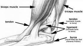
Injury to the muscle and tendons is called strain.

Reasons

  • Sudden unaccustomed or abrupt action or movements may tear the muscles.
  • Direct trauma can also injure the muscles and tendons.
  • Overstretching of muscles due to indirect trauma, especially in sports persons.

Types

  • Acute Strain: This is due to sudden violent force or direct trauma.
  • Chronic Strain: This is due to injury existing since a long period leading to muscle ischemia and fibrosis.

Path physiology

Injury to the muscles leads to pain. As a result, the muscle goes into spasm to limit the movements and reduce pain. Nevertheless, paradoxically, this protective muscle spasm causes pain due to stimulation of pain fibers and thus a vicious cycle sets. The painful stimuli cause muscle spasm through the peripheral nociceptive stimuli.

Severity of Strain

  • First Degree Strain (Mild Con Tusion)
  • This is due to blunt injury and is due to direct trauma of lose intensity.
  • Pathology: Few muscle fibers torn. Bleeding is minimal and the fascia remains intact.

Clinical Features

  • Localized pain and tenderness.
  • Pain and spasm prevents muscle stretching.
  • Function is not impaired largely.
  • Tenderness over the affected muscles.

Management:

  • First aid is by Cryotherapy (by application of ice) for a period of 20 minutes.
  • Gentle active muscle stretch may be permitted after 20 to 60 minutes.
  • Compression bandaging with optimum pressure.
  • Low dose and low power ultrasound helps.
  • Gentle massaging of the surrounding area helps.
  • If pain is minimal, the patient can be allowed to do the light work the next day.

Second Degree Strain

Cause: Here the trauma is more serious.

  • Pathology
  • Greater number of muscle fibers is torn.
  • There is bleeding.
  • The fascia is still intact.
  • Hematoma is still localized.

Symptoms: Here are the symptoms.

  • Pain is more severe.
  • Tenderness is severe.
  • Severe muscle spasm.
  • The patient is unable to move the limb.

Third Degree Strain

Cause: Undoubtedly, these injuries are due to trauma of a greater magnitude.

Pathology: Larger area and greater number of muscle fibers are involved. More than one muscle group may be involved. The fascia is partially torn.

Bleeding is widespread and more. There could be both intramuscular and inter muscular bleeding. The patient experiences severe pain and loss of function.

Symptoms: Here all the above symptoms are of greater intensity.

Treatment in Grade II and III Strains

  • For first 24 hours
  • Immediate application of ice.
  • Compression bandage.
  • Limb elevation.
  • Limb immobilized in splints.
  • Isometrics to the muscles, which are immobilized.
  • Active exercises to the unaffected joints.
  • Pulsed electromagnetic field therapy (PEMF) is known to help.
  • No active movements to the affected muscles.

During the Next 24 to 48 Hours

  • The pressure bandage is removed and active muscle exercises are begun.
  • Stretching within the limits of pain is commenced.
  • Thermotherapy: Ultrasound, short wave diathermy and TENS help to relieve pain.
  • Slow rhythmic massaging helps relieve the muscle
  • Non weight bearing on crutches is slowly started
  • Rest of the measures is the same as above.

Between 48 and 72 hours

Apart from all the measures mentioned so far, the additional measures during  this phase include:

  • More vigorous active movements are encouraged.
  • Deep transverse friction massage is added.
  • Partial weight bearing can be permitted.

After 72 hours

All the above measures are pursued in a more vigorous manner.

  • Pressure bandage is totally removed.
  • Progressive resisted exercises using the Fowler technique by taking out 10 to 12 repetition maximum (RM), is practiced.
  • Full weight bearing should be permitted in injuries of the lower limbs.
  • After full movement is regained, the patient is allowed to walk and jog.
  • Full functional activity should be regained by 4 to 6 weeks.
  • The various drugs used in the treatment of muscle strain to relieve pain and muscle stiffness is depicted.

Grade Four Strain

Cause: This is usually caused by severe trauma.

Pathology

  • Complete tear of the muscle.
  • The fascia is tom.
  • Considerable bleeding which is intramuscular and diffuse.
  • Gross swelling is present.

Clinical Features

  • Excruciating pain.
  • Severe tenderness is present.
  • A snapping sound may be heard by the patient.
  • Palpable gap between the muscles felt.
  • Severe loss of function.
  • Active movements produced by the agonist are absent.
  • Active muscle contraction is absent.
  • Joint function is not lost.
  • Muscle spasm is very severe.

Treatment

Surgery is advised.  This involves opening the ruptured site, evacuating the hematoma and suturing  the  fascia  sheath. Direct muscle repair is avoided.

  • Compression bandage is applied and the limb is immobilized for 2 to 3 weeks.
  • Active exercises to the unaffected joints.
  • Slow rhythmic isometric exercises to the affected muscles.
  • Non-weight bearing after 48 hours.
  • The use of low frequency current (faradism)  to obtain passive contraction  is very useful.
  • Deep heating modalities like ultrasound, etc. help.
  • Rest of the measures is same as for Grade II / III injuries.

If you suffer a muscle injury(Strains) which fails to respond after a few days or continues to niggle, please contact Alliance Physical Therapy for more specific advice. For more detailed Information Call Now at: 703-205-1919

http://www.alliancephysicaltherapyva.com/