Tag Archives: Physical Therapy

How to cure Ankle Tibial Nerve?

Clinical Features, Investigation and Treatment of Tibial Nerve:

images 22Stress fracture of the medial malleolus is an unusual injury but should be considered in the runner presenting with persistent medial ankle pain aggravated by activity. Although the fracture line is frequently vertical from the junction of the tibial plafond and the medial malleolus, it may arch obliquely from the junction to the distal tibial metaphysis.

Clinical Features

  • Athletes classically present with medial ankle pain that progressively increases with running and jumping activities.
  • Often they experience an acute episode, which leads to their seeking medical attention.
  • Examination reveals tenderness overlying the medial malleolus frequently in conjunction with an ankle effusion.

Investigations

  • In the early stages, X-rays may be normal, but with time a linear area of hyperlucency may be apparent, progressing to a lytic area and fracture line.
  • If the X-ray is normal, a radioisotopic bone scan, CT or MRI will be required to demonstrate the fracture.

Treatment

  • If no fracture or an undisplaced fracture is evident on X-ray, treatment requires weight-bearing rest with an air-cast brace until local tenderness resolves, a period of approximately six weeks.
  • If, however, a displaced fracture or a fracture that has progressed to non-union is present, surgery with internal fixation is required.
  • Following fracture healing, the practitioner should assess biomechanics and footwear. A graduated return to activity is required.

Stress bone injuries of the inside malleolus generally happen over time with extreme standing and walking action such as running. Physical rehabilitation treatment is essential for all sufferers with a stress crack of the inside malleolus to speed up treatment, avoid repeat and make sure an maximum result. At Alliance Physical Therapy we provide 24/7 access to online appointments, with most of the requests scheduled in less than 48 hours. For Best Rehabilitation and Physical Therapy Call now at: 703-750-1204

http://www.alliancephysicaltherapyva.com

How Foot and Ankle Discomfort is treated?

Causes and Treatment of Hallux Valgus:    Hallux valgus anatomy

Hallux Valgus is defined as a static subluxation of the first metatarsophalangeal joint. It is characterized by valgus (lateral) deviation of the great toe and varus (medial) deviation of the first metatarsal. Bony exostoses develop around the first metatarsophalangeal joint, often with an overlying bursitis. In severe cases, exostoses limit first metatarsophalangeal joint range of motion and cause pain with the pressure of footwear.

Causes

The development of hallux valgus appears to occur secondary to a combination of intrinsic and extrinsic causes. Recognized causative factors include:

  • Constricting footwear (e.g. high heels)
  • Excessive pronation-increased pressure on the medial border of the hallux, resulting in deformation of the medial capsular structures.
  • Others-cystic degeneration of the medial capsule, Achilles tendon contracture, neuro-muscular disorders, collagen deficient diseases.

Clinical Features

  • In the early phases hallux valgus is often asymptomatic, however, as the deformity develops, pain over the medial eminence occurs.
  • The pain is typicaIly relieved by removing the shoes or by wearing soft, flexible, wide-toed shoes. Blistering of the skin or development of an inflamed bursa over the medial eminence may occur.
  • In severe deformity, lateral metatarsalgia may occur due to the diminished weight-bearing capacity of the first ray.
  • Examination reveals the hallux valgus deformity often with a tender swelling overlying the medial eminence.

Investigation

Plain X-rays should be performed to assess both the severity of the deformity and the degree of first metatarsophalangeal joint degeneration.

Treatment

  • Initial treatment involves appropriate padding and footwear to reduce friction over the medial eminence.
  • Correction of foot function with orthoses is essential.
  • In more severe cases surgery may be required to reconstruct the first metatarsophalangeal joint and remove the bony exostoses.
  • Orthoses are often required after surgery.

Hallux valgus is a situation that impacts the combined at the platform of the big toe. This condition is commonly known as bunion. The big toe of the feet is known as the hallux. If the big toe begins to vary inward towards the child toe the situation is known as hallux valgus. Alliance Physical Therapy is best Rehab & Physical Therapy center in Virginia. Call now for quick Appointment: 703-704-5771

http://www.alliancephysicaltherapyva.com

How to get comfort from Thoracic Outlet Syndrome (Neck Tingling)?

Clinical Features and Treatment of Thoracic Outlet Syndrome:

Neck Tingling

The space at the thoracic outlet or inlet when it is less than adequate, subjects the neurovascular structures seeking to gain entry into the upper limbs via this space, to undue pressure. The blame for the neurovascular complaints should be placed at the doorstep of the decreased space and not at the structures producing the problems.

This syndrome results from the compression of neurovascular bundle comprising of subclavian artery and veinaxillary artery and vein and brachial plexus at the thoracic outlet. Thoracic outlet is a space between the first rib, clavicle, and the scalene muscles.

Sites of Compression

The sites of compression could be either Supraclavicular, Subclavicular or Infraclavicular.

  • Supraclavicular: Interscalene triangle between the anterior scalene muscles.
  • Subclavicular: Interval between the second thoracic rib, clavicle, and subclavius.
  • Infraclavicular: Beneath an enclosure formed by the coracoid process, pectoralis minor, and costocoracoid membrane.

Rare Cause

Scissor-like encirclement of axillary artery by the median nerve.

Contributing Factors

Dynamic Factors

Arm when in full abduction pulls up the artery by 180 degree causing compression in the short retroclavicular space.

Static Factors

  • Vigorous occupation: Increases the muscle bulk and thereby decreases the space.
  • Inactive occupation: Decreases the muscle bulk and thereby increases the space.
  • Congenital: Cervical rib decreases the interscalene space and thereby decreases the retroclavicular space.
  • Traumatic: Malunion or nonunion of fracture clavicle.

Anomalies of the first thoracic rib.

Miscellaneous

Clinical Features

Obviously, this syndrome poses two major problems. The first one relates to the compression of the major vessels and secondly to the compression of the nerves.

1.   Vascular Problems

Here the compression could be arterial or venous. During the arterial compression, which is mild in the early stages the patient complains of numbness of the whole arm with rapid fatigue during overhead exercises. If the compression is significant, the patient will complain of cold, cyanosis, pallor, and Raynaud’s phenomenon. Venous compression leaves the limb swollen and discolored after exercises, which disappears slowly with rest.

2.   Neurogenic Problems

Patients complain of par esthesia along the medial aspect of the arm, hand, little and ring fingers. There is weakness of the hand also.

Complications

  • Subclavian artery compression
  • Results in poststenotic dilatation
  • Stasis favors thrombosis
  • The thrombi break and migrate distally causing embolization

Investigations

  • Nerve Conduction Studies: Difficult to determine the nerve conduction velocity through the thoracic outlet, but its biggest value is to rule-out problems like entrapment, e.g. ulnar nerve at elbowwrist, etc.

Treatment

  • Conservative treatment: Consists of rest, physiotherapy, exercises like shoulder shrugging, etc.
  • Surgical treatment

Thoracic Outlet problem is a number of conditions that happen when the veins or anxiety in the thoracic store — the area between your collarbone and your first rib — become compacted. This can cause discomfort in shoulder area and throat and pins and needles in your fingertips. Call now for best Physical Therapy: 703-205-1919

http://www.alliancephysicaltherapyva.com

How we treat Fracture of Femur?

Classification And Treatment in Supracondylar Fracture of Femur:SUPRACONDYLAR FRACTURE OF FEMUR

Supracondylar region extends from the femoral condyles to the junction of metaphysis with femoral shaft .The distal fragment is displaced and angulated posteriorly due to the pull of gastrocnemius muscle.

Mechanism of Injury

It is due to severe valgus or varus forces with axial loading and rotation due to RTA, fall, etc.

Classification

  1. Nears’s Classification
  • Undisplaced Fracture
  • Displaced Fracture
  • Medial Displacement
  • Lateral Displacement
  • Comminuted Fracture
  1. Muller’s AO Classification
  • Type A: Extra-articular Fractures.
  • Type B: Unicondylar Fractures.
  • Type C: Bicondylar Fractures.

Each is further subdivided into 1-3 depending on the severity of comminution.

  1. OTA Classification of Supracondylar Fractures of Femur

Supracondylar Fractures of Femur

  • Type A: Extra-articular.
  • Type B: Partial articular (Unicondylar).
  • Type C: Total articular (Bicondylar).
  1. Further Subdivisions

Type A

Type B

Type C

  • Articular and metaphyseal simple.
  • Articular simple and metaphyseal comminution.
  • Total comminution.

Clinical Features

It consists of the usual features of fractures, but what is specific to this fracture is the flexion deformity caused by the pull of gastrocnemius. Hemarthrosis is commonly seen, especially with fractures extending into the joint.

Radiographs

Radiograph helps to study the fracture pattern more accurately. Routine AP, lateral and oblique (45degree) views are required.

Arteriography: This should be performed in suspected vascular damage or in associated dislocation of the knee joint.

Treatment

The treatment usually consists of conservative methods, traction and operative methods.

  • Conservative Methods: This has a limited role and is usually useful in impacted and undisplaced fractures. In the former, a long leg or Spica cast is sufficient and in the latter, a long above knee cast after an initial period of skin or skeletal traction is all that is required.
  • Traction Methods: The choice is mainly skeletal traction and two methods are described.
  • Upper Tibial Traction: Here the skeletal traction is applied through the upper end of tibia. Initial weight used is around 15-20 lbs and is subsequently reduced. The traction is given for a period of 8-12 weeks and the patient is put on cast braces. To prevent the knee stiffness from developing, the patient is encouraged to carry out the knee movements during the traction itself.
  • Two-Pin Traction Method: In this method, traction is added through the distal femur apart from the traction given through the upper end of tibia. This helps in accurate reduction of the fracture and maintains the reduction so obtained. The disadvantage of this technique is that it is cumbersome and may cause neurovascular compressions in and around the knee.
  • Operative Methods: This consists of DRIP and is preferred as the closed reduction is associated with troublesome complications like limited knee motion, residual varus and internal rotation deformities. The advantages of open reduction are early mobilization of the knee joint and an accurate reduction and rigid fixation.
  • Fixation Methods: The choice is between medullary fixation and blade plate fixation.
  • Intramedullary Fixations: Rush pins, Ender’s nail, medullary nails, split nails, static locking nails, etc. are some of the commonly used medullary fixation methods. They offer biological fixation but the fixation offered is less stable.
  • Trigen (Third generation) Knee Nail: Inserted in a retrograde fashion. It is a titanium nail and has two holes for oblique screws and one for transverse screw at the insertion end. At the opposite locking end two holes are present in the anteroposterior plane and 2 holes in the lateral plane. The results are encouraging.

Complications

The complications commonly encountered in supracondylar fractures are delayed union, mal union, nonunion, injury to the popliteal vessels and common peroneal nerves, knee stiffness, deep vein thrombosis, infection, implant failure, etc.

If you are being affected bone fracture of femur and come instantly our clinic Alliance Physical Therapy. Our highly skilled hand therapists are proficient in the treatment of hand and upper extremity pathologies, from the acute to the chronic. Under the direction of the referring physician, our team designs each treatment based on the physician’s diagnosis and the specific needs of the patient. Conditions treated include, but are not limited to arthritis, fractures, tendon injuries, peripheral nerve injuries, carpal tunnel syndrome, crush injuries and repetitive motion disorder. For more detailed information our clinic Call Now at: 703-205-1919

http://www.alliancephysicaltherapyva.com/

How to Care for Muscle Injury Pain?

Types, Causes, Symptoms And Treatment of Muscle Injuries (Strain):Muscle Injury (Strains)

Definition of Muscle Injury:

Injury to the muscle and tendons is called strain.

Reasons

  • Sudden unaccustomed or abrupt action or movements may tear the muscles.
  • Direct trauma can also injure the muscles and tendons.
  • Overstretching of muscles due to indirect trauma, especially in sports persons.

Types

  • Acute Strain: This is due to sudden violent force or direct trauma.
  • Chronic Strain: This is due to injury existing since a long period leading to muscle ischemia and fibrosis.

Path physiology

Injury to the muscles leads to pain. As a result, the muscle goes into spasm to limit the movements and reduce pain. Nevertheless, paradoxically, this protective muscle spasm causes pain due to stimulation of pain fibers and thus a vicious cycle sets. The painful stimuli cause muscle spasm through the peripheral nociceptive stimuli.

Severity of Strain

  • First Degree Strain (Mild Con Tusion)
  • This is due to blunt injury and is due to direct trauma of lose intensity.
  • Pathology: Few muscle fibers torn. Bleeding is minimal and the fascia remains intact.

Clinical Features

  • Localized pain and tenderness.
  • Pain and spasm prevents muscle stretching.
  • Function is not impaired largely.
  • Tenderness over the affected muscles.

Management:

  • First aid is by Cryotherapy (by application of ice) for a period of 20 minutes.
  • Gentle active muscle stretch may be permitted after 20 to 60 minutes.
  • Compression bandaging with optimum pressure.
  • Low dose and low power ultrasound helps.
  • Gentle massaging of the surrounding area helps.
  • If pain is minimal, the patient can be allowed to do the light work the next day.

Second Degree Strain

Cause: Here the trauma is more serious.

  • Pathology
  • Greater number of muscle fibers is torn.
  • There is bleeding.
  • The fascia is still intact.
  • Hematoma is still localized.

Symptoms: Here are the symptoms.

  • Pain is more severe.
  • Tenderness is severe.
  • Severe muscle spasm.
  • The patient is unable to move the limb.

Third Degree Strain

Cause: Undoubtedly, these injuries are due to trauma of a greater magnitude.

Pathology: Larger area and greater number of muscle fibers are involved. More than one muscle group may be involved. The fascia is partially torn.

Bleeding is widespread and more. There could be both intramuscular and inter muscular bleeding. The patient experiences severe pain and loss of function.

Symptoms: Here all the above symptoms are of greater intensity.

Treatment in Grade II and III Strains

  • For first 24 hours
  • Immediate application of ice.
  • Compression bandage.
  • Limb elevation.
  • Limb immobilized in splints.
  • Isometrics to the muscles, which are immobilized.
  • Active exercises to the unaffected joints.
  • Pulsed electromagnetic field therapy (PEMF) is known to help.
  • No active movements to the affected muscles.

During the Next 24 to 48 Hours

  • The pressure bandage is removed and active muscle exercises are begun.
  • Stretching within the limits of pain is commenced.
  • Thermotherapy: Ultrasound, short wave diathermy and TENS help to relieve pain.
  • Slow rhythmic massaging helps relieve the muscle
  • Non weight bearing on crutches is slowly started
  • Rest of the measures is the same as above.

Between 48 and 72 hours

Apart from all the measures mentioned so far, the additional measures during  this phase include:

  • More vigorous active movements are encouraged.
  • Deep transverse friction massage is added.
  • Partial weight bearing can be permitted.

After 72 hours

All the above measures are pursued in a more vigorous manner.

  • Pressure bandage is totally removed.
  • Progressive resisted exercises using the Fowler technique by taking out 10 to 12 repetition maximum (RM), is practiced.
  • Full weight bearing should be permitted in injuries of the lower limbs.
  • After full movement is regained, the patient is allowed to walk and jog.
  • Full functional activity should be regained by 4 to 6 weeks.
  • The various drugs used in the treatment of muscle strain to relieve pain and muscle stiffness is depicted.

Grade Four Strain

Cause: This is usually caused by severe trauma.

Pathology

  • Complete tear of the muscle.
  • The fascia is tom.
  • Considerable bleeding which is intramuscular and diffuse.
  • Gross swelling is present.

Clinical Features

  • Excruciating pain.
  • Severe tenderness is present.
  • A snapping sound may be heard by the patient.
  • Palpable gap between the muscles felt.
  • Severe loss of function.
  • Active movements produced by the agonist are absent.
  • Active muscle contraction is absent.
  • Joint function is not lost.
  • Muscle spasm is very severe.

Treatment

Surgery is advised.  This involves opening the ruptured site, evacuating the hematoma and suturing  the  fascia  sheath. Direct muscle repair is avoided.

  • Compression bandage is applied and the limb is immobilized for 2 to 3 weeks.
  • Active exercises to the unaffected joints.
  • Slow rhythmic isometric exercises to the affected muscles.
  • Non-weight bearing after 48 hours.
  • The use of low frequency current (faradism)  to obtain passive contraction  is very useful.
  • Deep heating modalities like ultrasound, etc. help.
  • Rest of the measures is same as for Grade II / III injuries.

If you suffer a muscle injury(Strains) which fails to respond after a few days or continues to niggle, please contact Alliance Physical Therapy for more specific advice. For more detailed Information Call Now at: 703-205-1919

http://www.alliancephysicaltherapyva.com/

How to treat Longstanding Groin Pain?

Treatment of Adductor-Related Longstanding Groin Pain:    Adductor Muscle Strain

Longstanding adductor-related groin pain is localized medially in the groin and may radiate down along the adductor muscles. The key examination features that distinguish this clinical entity from others are maximal tenderness at the adductor tendon insertion and pain with resisted adduction. Weakness of the adductor muscles is common and palpation of the adductors reveals generally increased muscle tone with trigger points along the adductor longus. The pubic symphysis is frequently tender but this does not help to differentiate the four clinical entities.

Occasionally there may be an obvious adductor tendinopathy or enthesopathy with localized tenderness, pain, and weakness on contraction, especially eccentric contraction, and a typical appearance of tendinopathy on ultrasound or MRI examination. More frequently there is no specific tendinopathy present.

Signs and Symptoms of Groin Pain

Unfortunately most patients with adductor-related groin pain continue to train and play until pain prevents them from running. When the condition has reached that stage, a lengthy period of rest and rehabilitation is usually required. However, if early warning signs are heeded, appropriate measures may prevent the development of the full blown syndrome. These early clinical warning signs are (from most common to least):

  • Tightness/stiffness during or after activity with nil (or temporary only) relief from stretching
  • Loss of acceleration
  • Loss of maximal sprinting speed
  • Loss of distance with long kick on run
  • Vague discomfort with deceleration.

Treatment of Groin Pain

Traditional treatment for most types of groin pain was ‘rest’ but this most often resulted in a return of symptoms on resumption of activity. Compared with rest and passive electrotherapy, active rehabilitation provides more than 10 times the likelihood of pain-free successful return to sport. Thus, we outline a treatment protocol that combines experience and evidence from leading clinical centers. Five basic principles underpin a treatment regimen:

  • Ensure that exercise is performed without pain.
  • Identify and reduce the sources of increased load on the pelvis.
  • Improve lumbopelvic stability.
  • Strengthen local musculature using proven protocols.
  • Progress the patient’s level of activity on the basis of regular clinical assessment.

These are outlined below.

1.   Ensure that exercise is performed without pain

The first and most important step is for the patient to cease training and playing in pain. Pain-free exercise is absolutely crucial for this rehabilitation program. If pain is experienced during any of the rehabilitation activities, or after them, that activity should be reduced or ceased altogether. Experienced clinicians use absence of pain on the key provocation tests (e.g. squeeze test and Thomas test) as a guide to progress the rehabilitation program and minimize the mechanical stress on injured tissues.

2.   Identify and reduce the sources of increased load on the pelvis

As discussed previously, it is essential to identify and reduce the sources of increased load on the pubic bones. This may involve:

  • Reducing adductor muscle tone and guarding with soft tissue treatment and/or dry needling
  • Correcting iliopsoas muscle shortening with local soft tissue treatment, neural stretching and mobilization of upper lumbar intervertebral joints
  • Reducing glutens medius muscle tone and myofascial shortening with soft tissue treatment and/or dry needling
  • Identifying and correcting any hip joint abnormality
  • Mobilizing stiff intervertebral segments
  • Improving core stability, especially activation of transversus abdominis and anterior pelvic floor muscles.

3.   Improve lumbopelvic stability

Research has demonstrated a delayed onset of action of transversus abdominis activity in patients with longstanding groin pain, suggesting that impaired core or lumbopelvic stability plays a role in the development of this condition.

4.   Strengthen local musculature using proven protocols

Once pain has settled and muscle shortening has been corrected in the adductor, iliopsoas and gluteal muscles, then a graduated pain-free muscle strengthening program can be commenced. A similar pre-season adductor muscle strengthening program reduced the incidence of adductor muscle strains in ice hockey players who were identified as at risk.

5.   Progress the patient’s level of activity on the basis of regular clinical assessment

The aim of the graded exercise program is to gradually increase the load on the pubic bones and surrounding tissues. Once the patient is pain-free, pain-free walking can begin and be gradually increased in speed and distance. The criteria for when the patient may return to running are when:

  • Brisk walking is pain-free
  • Resisted hip flexion in the Thomas position is pain-free
  • There is no ‘crossover’ sign
  • There is minimal adductor guarding.

Other non-surgical treatments

  • Compression shorts have been advocated for those with mild pain who insist on continuing to train and play, and for those returning to sport after rehabilitation.
  • The shorts substantially reduced pain when worn during exercise.
  • The mechanism of action of compression shorts remains unclear, but Dutch researchers have reported that groin pain on resisted adduction (the ‘squeeze test’) was significantly reduced by the application of a pelvic belt.
  • They speculated that relative pelvic instability may contribute to the groin pain typically attributed to tendinopathy.

Groin Pain is very common among athletes. A significant cause of long-standing issues is adductor-related groin discomfort. Alliance Rehab and Physical Therapy provide 24/7 access to online appointments. If you are suffering from Groin Pain then Contact us at our website or Call us at: 703-750-1204

http://www.alliancephysicaltherapyva.com

How to Prevent Patella Fracture?

Clinical Features and Treatment of Acute Patellar (Kneecap) Trauma: Patella Injury

Acute trauma to the patella (e.g. from a hockey stick or from a fall onto the kneecap) can cause a range of injuries from fracture of the patella to osteochondral damage of the patellofemoral joint with persisting patellofemoral joint pain. In some athletes, the pain settles without any long-term sequelae. If there is suspicion of fracture, X-ray should be obtained. It is important to be able to differentiate between a fracture of the patella and a bipartite patella. A skyline view of the patella should be performed in addition to normal views. If there is no evidence of fracture, the patient can be assumed to be suffering acute patellofemoral inflammation. This can be a difficult condition to treat. Treatment consists of NSAIDs, local electrotherapy (e.g. interferential stimulation, TENS) and avoidance of aggravating activities such as squatting or walking down stairs. Taping of the patella may alter the mechanics of patellar tracking and therefore reduce the irritation and pain.

Fracture of the Patella

  • Patellar fractures can occur either by direct trauma, in which case the surrounding retinaculum can be intact, or by indirect injury from quadriceps contraction, in which case the retinaculum and the vastus muscles are usually torn.
  • Undisplaced fractures of the patella with normal continuity of the extensor mechanism can be managed conservatively, initially with an extension splint.
  • Over the next weeks as the fracture unites, the range of flexion can be gradually increased and the quadriceps strengthened in the inner range.
  • Fractures with significant displacement, where the extensor mechanism is not intact, require surgical treatment. This involves reduction of the patella and fixation, usually with a tension band wire technique.
  • The vastus muscle on both sides also needs to be repaired. The rehabilitation following this procedure is as for undisplaced fracture.

Patella Dislocation

  • Patella dislocation occurs when the patella moves out of its groove laterally onto the lateral femoral condyle.
  • Acute patella dislocation may be either traumatic with a good history of trauma and development of a hemarthrosis following injury, or atraumatic, which usually occurs in young girls with associated ligamentous laxity, does not have a good history of trauma, and is accompanied by mild-to-moderate swelling.

Clinical Features

Patients with traumatic patella dislocation usually complain that, on twisting or jumping, the knee suddenly gave way with the development of severe pain. Often the patient will describe a feeling of something ‘popping out’. Swelling develops almost immediately. The dislocation usually reduces spontaneously with knee extension; however, in some cases this may require some assistance or regional anesthesia (e.g. femoral nerve block). A number of factors predispose to dislocation of the patella:

  • Femoral anteversion
  • Shallow femoral groove
  • Genu valgum
  • Loose medial retinaculum
  • Tight lateral retinaculum
  • Vastus medialis dysplasia
  • Increased Q angle
  • Patellar alta
  • Excessive subtalar pronation
  • Patellar dysplasia
  • General hypermobility

The main differential diagnosis of patella dislocation is an ACL rupture. Both conditions have similar histories of twisting, an audible ‘pop’, a feeling of something ‘going out’ and subsequent development of hemarthrosis. On examination, there is usually a gross effusion marked tenderness over the medial border of the patella and a positive lateral apprehension test when attempts are made to push the patella in a lateral direction. Any attempt to contract the quadriceps muscle aggravates the pain. X-rays, including anteroposterior, lateral, skyline, and intercondylar views, should be performed to rule out osteochondral fracture or a loose body.

Treatment

  • Treatment of traumatic patella dislocation depends on presentation. Relatively atraumatic dislocations are treated conservatively.
  • Traumatic first- or second-time dislocations (hemarthrosis present) are treated with arthroscopic washout and debridement.
  • Recurrent dislocation is treated with surgical stabilization.
  • As a result, the rehabilitation program is lengthy and emphasizes core stability, pelvic positioning, vastus medialis obliquus strength, and stretching of the lateral structures when tight.
  • The most helpful addition to patellofemoral rehabilitation in the recent past is increased emphasis on core stability.
  • Similar to ACL intervention exercises, rotational control of the limb under the pelvis is critical to knee and kneecap stability.

Patella Fracture is common among athletes. It is an injury to kneecap. Major symptom of Patella Fracture is knee swelling. Alliance Rehab & Physical Therapy is the best Rehab & Physical Therapy center in Virginia. We provide 24/7 access to online appointments, with most of the requests scheduled in less than 48 hours.

Contact Us at: 703-751-1008

http://www.alliancephysicaltherapyva.com

Are you being affected by Lower Back Pain?

Spondylolisthesis: Back Condition And Treatment

Spondylolisthesis: Back Condition And Treatment

Spondylolisthesis refers to the slipping of part or all of one vertebra forward on another. The term is derived from the Greek spondylos, meaning vertebra and olisthanein, meaning to slip or slide down a slippery path.

It is often associated with bilateral pars defects that usually develop in early childhood and have a definite family predisposition. Pars defects that develop due to athletic activity (stress fractures) rarely result in spondylolisthesis.

Spondylolisthesis is most commonly seen in children between the ages of 9 and 14, in the vast majority of cases it is the LS vertebra that slips forward on the S1. The spondylolisthesis is graded according to the degree of slip of the vertebra. A grade I slip denotes that a vertebra has slipped up to 25% over the body of the vertebra underlying it; in a grade II slip the displacement is greater than 25%; in a grade III slip, greater than 50%; and in a grade IV slip, greater than 75%. Lateral X-rays best demonstrate the extent of vertebral slippage.

Clinical Features

Grade I spondylolisthesis is often asymptomatic and the patients may be unaware of the defect. Patients with grade II or higher slips may complain of low back pain, with or without leg pain. The back pain is aggravated by extension activities.

On examination, there may be a palpable dip corresponding to the slip. Associated soft tissue abnormalities may be present. In considering the treatment of this condition, it is important to remember that the patient’s low back pain is not necessarily being caused by the spondylolisthesis.

Treatment

Treatment of athletes with grade I or grade II symptomatic spondylolisthesis involves:

  • Rest from aggravating activities combined with abdominal and extensor stabilizing exercises and hamstring stretching.
  • Antilordotic bracing, which may also be helpful.
  • Mobilization of stiff joints above or below the slip on clinical assessment; gentle rotations may be helpful in reducing pain; manipulation should not be performed at the level of the slip.
  • Athletes with grade I or grade II spondylolisthesis may return to sport after treatment when they are pain free on extension and have good spinal stabilization.
  •  If the symptoms recur, activity must be ceased.
  • Athletes with grade III or grade IV spondylolisthesis should avoid high speed or contact sports.
  • Treatment is symptomatic. It is rare for a slip to progress; however, if there is evidence of progression, spinal fusion should be performed.

If you are being impacted in low back pain again then come instantly at Alliance Physical Therapy Center in VA & DC. Our Reduced Lower back Program uses a consistent, functional and outcomes-oriented approach to care that concentrates specifically on the lower back. Through an active and educational procession of treatment, our practitioners assist the affected person in returning to normal, activities as soon as possible. By providing comprehensive education in structure, pathology and proper proper the lower back, the affected person is motivated to participate in his or her recovery and in the prevention of future injury. For more information Call Now at: 703-205-1919

http://www.alliancephysicaltherapyva.com/

How we get relief from Minimizing Extent of Injury (RICE)?

Meaning of R.I.C.E.

The most important time in the treatment of acute soft tissue injuries is in the 24 hours immediately following injury. When soft tissue is injured, blood vessels are usually damaged too.

Thus, blood accumulates around damaged tissue and compresses adjoining tissues, which causes secondary hypoxic injury and further tissue damage. Consequently, every effort should be made to reduce bleeding at the site of injury. The most appropriate method of doing this is summarized by the letters RICE.

  • R – Rest
  • I -  Ice
  • C – Compression
  • E – Elevation

Rest

Whenever possible following injury the athlete should cease activity to decrease bleeding and swelling. For example, with a thigh contusion, bleeding will be increased by contraction of the quadriceps muscle during running. Where necessary, complete rest can be achieved with the use of crutches for a lower limb injury or a sling for upper limb injuries.

Ice

Immediately after injury, ice is principally used to reduce tissue metabolism. Ice is also used in the later stages of injury treatment as a therapeutic modality.

Ice can be applied in a number of forms:

  • Crushed ice can be wrapped in a moist cloth or towel and placed around the injured area, held in place with a crepe bandage.
  • Reusable frozen gel packs.
  • Instant ice packs that do not need pre-cooling.
  • Ice immersion in a bucket (useful for treatment of injuries of the extremities).
  • Cold water and cooling sprays, which are often used in the immediate treatment of injuries but are unlikely to affect deeper tissues.

Although there is no high-quality evidence for how long, and how often, to apply ice after an acute injury, a systematic review suggested that intermittent 10-minute ice treatments are most effective at cooling injured animal tissue and healthy human tissue. Many practitioners apply ice for 15 minutes every I -2 hours initially and then gradually reduce the frequency of application over the next 24 hours.

Ice should not be applied where local tissue circulation is impaired (e.g. in Raynaud’s phenomenon, peripheral vascular disease) or to patients who suffer from a cold allergy. Other adverse effects of prolonged ice application are skin burns and nerve damage.

Compression

Compression of the injured area with a firm bandage reduces bleeding and, therefore, minimizes swelling. Compression should be applied both during and after ice application; the width of the bandage applied varies according to the injured area.

 The bandage should be applied firmly but not so tightly as to cause pain. Bandaging should start just distal to the site of bleeding with each layer of the bandage overlapping the underlying layer by one-half. It should extend to at least a hands breadth proximal to the injury margin.

Elevation

Elevation of the injured part decreases hydrostatic pressure and, thus, reduces the accumulation of interstitial fluid. Elevation can be achieved by using a sling for upper limb injuries and by resting lower limbs on a chair, pillows or bucket. It is important to ensure that the lower limb is above the level of the pelvis.

Other minimizing factors

In the initial phase of injury (first 24 hours), heat and heat rubs, alcohol, moderate/intense activity and vigorous soft tissue therapy should all be avoided? Whether or not electrotherapeutic modalities (e.g. magnetic field therapy, interferential stimulation, TENS) provide effective pain relief and reduction of swelling in the initial period is a subject of debate.

It is usually suggested to exercise R.I.C.E. at duration of 4 to 6 time for up to 48 time after an damage. Heat therapies are appropriate for some accidents, but should only be regarded after swelling has receded, roughly 72 time after an damage. If the part of one’s body does not reply to R.I.C.E. treatment within 48 time, it would be sensible to seek advice from your doctor in the occasion a serious damage has happened such as inner blood loss or a damaged cuboid.

At Alliance Physical Therapy Our team works with individuals who have undergone a total hip or knee replacement with arthroscopic or other surgeries, sustained trauma to a bone, or have a bone or soft tissue disease. We focus on helping patients regain their strength, mobility and endurance so they can return home and resume their regular routines.For More Information Call At: 703-205-1919

http://www.alliancephysicaltherapyva.com/

How to Recognize a Condition Masquerading as a Sports Injury?

Conditions Masquerading as Sports Injury:

Not every patient who presents to the sports medicine clinician has a sports-related condition. Sports medicine, like every branch of medicine, has its share of conditions that must not be missed but appear at first to be rather benign conditions. The aim of this article is to remind you that the patient with the minor ‘calf strain‘ may, in fact, have a deep venous thrombosis, or that the young basketball player who has been labeled as having Osgood – Schlatter disease because of  playing may actually have an osteosarcoma.  The first part of the article outlines a clinical approach that should maximize your chances of recognizing a condition that is masquerading as a sports-related condition. The second part of the article describes some of these conditions and illustrates how they can present in the sports medicine setting.

Examination

The key to recognizing that everything is not as the first impression might suggest is to take a thorough history and perform a detailed physical examination. If the clinician has not recognized a masquerading condition from the history and examination, it is unlikely that he or she will order the appropriate investigations to make the diagnosis. For example, if a patient presents with tibial pain and it is, in fact, due to hypocalcaemia secondary to lung cancer, a bone scan of the tibia looking for stress fracture will usually not help with the diagnosis, but a history of weight loss, occasional hemoptysis and associated shoulder pain, the history of associated arm tightness and the physical finding of prominent superficial veins are more important clues to axillary vein thrombosis than would be a gray-scale ultrasound scan looking for rotator cuff tendinopathy.

If there is something about the history and examination that does not fit the pattern of the common conditions, then consider alternative, less common conditions. To be able to make the diagnosis of a rare or non-musculoskeletal condition, you must ask yourself, Could this be a rare condition or unusual manifestation? Then other options are entertained, and the appropriate diagnosis can be conceived. Thus, successful diagnosis of masquerading conditions requires recognition of a discrepancy between the patient’s clinical features and the typical pattern that one is familiar with from clinical experience.

Bone and Soft Tissue Tumors

Primary malignant tumors of bone and soft tissues are rare but when they occur it is most likely to be in the younger age group (second to third decade). Osteosarcomata can present at the distal or proximal end of long bones, more commonly in the lower limb, producing joint pain. Patients often recognize that pain is aggravated by activity and hence present to the sports medicine clinic. The pathological diagnosis of osteosarcoma is dependent on the detection of tumor producing bone and so an X-ray may reveal a moth eaten appearance with new bone formation in the soft tissues and lifting of the periosteum (Codman’s triangle) .In young patients, the differential diagnosis includes osteomyelitis. It is recommended that any child or adolescent with bone pain be X-rayed. Surgery is the preferred treatment.

Synovial sarcomata frequently involve the larger lower joints such as the knee and ankle. Patients present with pain, often at night or with activity, maybe with instability and swelling.

Synovial chondromatosis and pigmented villonodular synovitis are benign tumors of the synovium found mainly in the knee, which present with mechanical symptoms.

Osteoid osteoma is a benign bone tumor that often presents as exercise-related bone pain and tenderness and is, therefore, frequently misdiagnosed as a stress fracture. The bone scan appearance is also similar to that of a stress fracture, although the isotope uptake is more intense and widespread. This condition is characterized clinically by the presence of night pain and by the abolition of symptoms with the use of aspirin. The tumor has a characteristic appearance on CT scan with a central nidus.

Ganglion cysts are lined by connective tissue, contain mucinous fluid and are found mainly around the wrist, hand, knee and foot. They may be to a joint capsule or tendon sheath and may have a connection to the synovial cavity. They are usually asymptomatic but can occasionally cause pain and deformity.

Rheumatological Conditions

These are dealt with in greater detail in the section on multiple joint problems. Patients with inflammatory musculoskeletal disorders frequently present to the sports medicine clinic with a masquerading traumatic or mechanical condition. Low back pain of ankylosing spondylitis, psoriatic enthesopathy or early rheumatoid arthritis is common examples.

In patients presenting with an acutely swollen knee without a history of precipitant trauma or patellar tendinopathy without overuse, the clinician may be alerted to the possibility that these are inflammatory in origin. Prominent morning joint or back stiffness, night pain or extra-articular manifestations of rheumatologlcal conditions (e.g. skin rashes, nail abnormalities), bowel disturbance, eye involvement (conjunctivitis, iritis) or urethral discharge may all provide clues.

Disorders of Muscle

Dermotomyositis and polymyositis are inflammatory connective tissue disorders characterized by proximal limb girdle weakness, often without pain Dermatomyositis, unlike polymyositis, is also associated with a photosensitive skin rash in light-exposed areas (hands and face). In the older adult, dermatomyositis may be associated with malignancy in approximately 50% of cases. The primary malignancy may be easily detectable or occult. In the younger adult, weakness may be profound (e.g. unable to rise from the floor) but in the early stages may manifest only as under-performance in training or competition.

Dermatomyositis and polymyositis may also be associated with other connective tissue disorders such as systemic lupus erythematosus or systemic sclerosis, and muscle abnormality is characterized by elevated creatine kinase levels and electromyography (EMG) and muscle biopsy changes.

Regional dystrophies such as limb girdle dystrophy and facio-scapulo-humeral dystrophy may also adults. They are also associated with characteristic changes.

Endocrine Disorders

Several endocrine disorders, for example, hypothyroidism and hyperparathyroidism, may be associated with the deposition of calcium pyrophosphate in joints. Patients may develop acute pseudo gout or a polyarticular inflammatory arthritis resembling rheumatoid arthritis. X-rays of the wrists or knees may demonstrate chondrocalcinosis of the menisci or triangular fibro cartilage complex. Adhesive capsulitis or septic arthritis may be the presenting complaint in patients with diabetes mellitus and those with other endocrine disorders such as acromegaly may develop premature osteoarthritis or carpal tunnel syndrome. Patients with hypocalcaemia secondary to malignancy (e.g. of the lung) or other conditions such as hyperparathyroidism can present with bone pain as well as constipation, confusion and renal calculi. A proximal myopathy may develop in patients with primary Cushings syndrome or after corticosteroid use.

Vascular Disorders

Patients with venous thrombosis or arterial abnormalities may present with limb pain and swelling aggravated by exercise. Calf, femoral or axillary veins are common sites for thrombosis. While a precipitant cause may be apparent (e.g. recent surgery or air travel), consider also the thrombophilias such as the antiphospholipid syndrome or deficiencies of protein C, protein S, anti thrombin III or factor V Leiden.

The Claudicant pain of peripheral vascular disease is most likely to be first noticed with exercise and so patients may present to the sports medicine practioner. Remember also that arteriopathy can occur in patients with diabetes. Various specific vascular entrapments are also found, such as popliteal artery entrapment, which presents as exercise related calf pain, and thoracic outlet syndrome.

Genetic Disorders

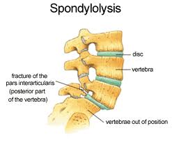
Marfans syndrome is an autosomal dominant disorder of fibril in characterized by musculoskeletal, cardiac and ocular abnormalities. Musculoskeletal problems are common due to joint hyper mobility, ligament laxity, scoliosis or spondylolysis. In patients with the Marfanoid habitus, referral for echocardiography and ophthalmological opinion should be considered as sudden carac death or lens dislocation may result. Hemochromatosis is an autosomal recessive disorder of iron handling, which results in iron overload.

Patients may present with a calcium pyrophosphate arthropathy with characteristic involvement of the second and third metacarpophalangeal joints and hook-shaped osteophytes seen on X-ray of these joints. While ferritin levels are raised in patients with hemochromatosis, it is important to remember that ferritin is also an acute-phase protein and so levels can be elevated in response to inflammatory arthropathy.

Infection

Bone and joint infections, while uncommon, may have disastrous consequences if the diagnosis is missed. Bone pain in children, worse at night or with activity, should alert the clinician to the possibility of osteomyelitis. Bone infection near a joint may result in a reactive joint effusion. Septic arthritis is rare in the normal joint. In arthritic, recently arthrocentesed or diabetic joints, sepsis is much more common. Rapid joint destruction may follow if left untreated.

Even though Staphylococcus aureus is the causative organism in more than 50% of cases of acute septic joints, it is imperative that joint aspiration for Gram stain and culture and blood cultures are taken before commencement of antibiotic treatment. Once only or repeated joint lavage may be considered in patients receiving intravenous antibiotic treatment. The immune compromised patient may present with a chronic septic arthritis. In this situation, tuberculosis or fungal infections should be considered.

Regional Pain Syndromes

Complex regional pain syndrome type 1 (formerly known as reflex sympathetic dystrophy [RSD]) is a post-traumatic phenomenon characterized by localized pain out of proportion to the injury, vasomotor disturbances, edema and delayed recovery from injury. The vasomotor disturbances of an extremity manifest as vasodilatation (warmth, redness) or vasoconstriction (coolness, cyanosis, mottling). Early mobilization and avoidance of surgery are two important keys to successful management.

Myofascial pain syndromes develop secondary to either acute or overuse trauma. They present as regional pain associated with the presence of one or more active trigger points.

Fibromyalgia is a chronic pain syndrome characterized by widespread pain, chronic fatigue, decreased pain threshold, sleep disturbance, psychological stress and diffusely tender muscles. It is often associated with other symptoms, including irritable bowel syndrome, dyspareunia, headache, irritable bladder and subjective joint swelling and pain. Fibromyalgia is diagnosed on the examination finding of 11of 18 specific tender point sites in a patient with widespread pain. Current treatment evidence is for a stepwise program emphasizing education, certain medications, exercise and cognitive therapy. Chronic fatigue syndrome has many similarities to fibromyalgia’s and may be the same disease process. It may present as excessive post-exercise muscle soreness but is always associated with excessive fatigue. Behavioral therapy and graded exercise therapy have shown promise as treatment.

At Alliance Physical Therapy we provide 24/7 access to online appointments, with most of the requests scheduled in less than 48 hours. For More Information Call Now at: 703-751-1008

http://www.alliancephysicaltherapyva.com/